• How Long Is Hand Foot Mouth Contagious and When to Return

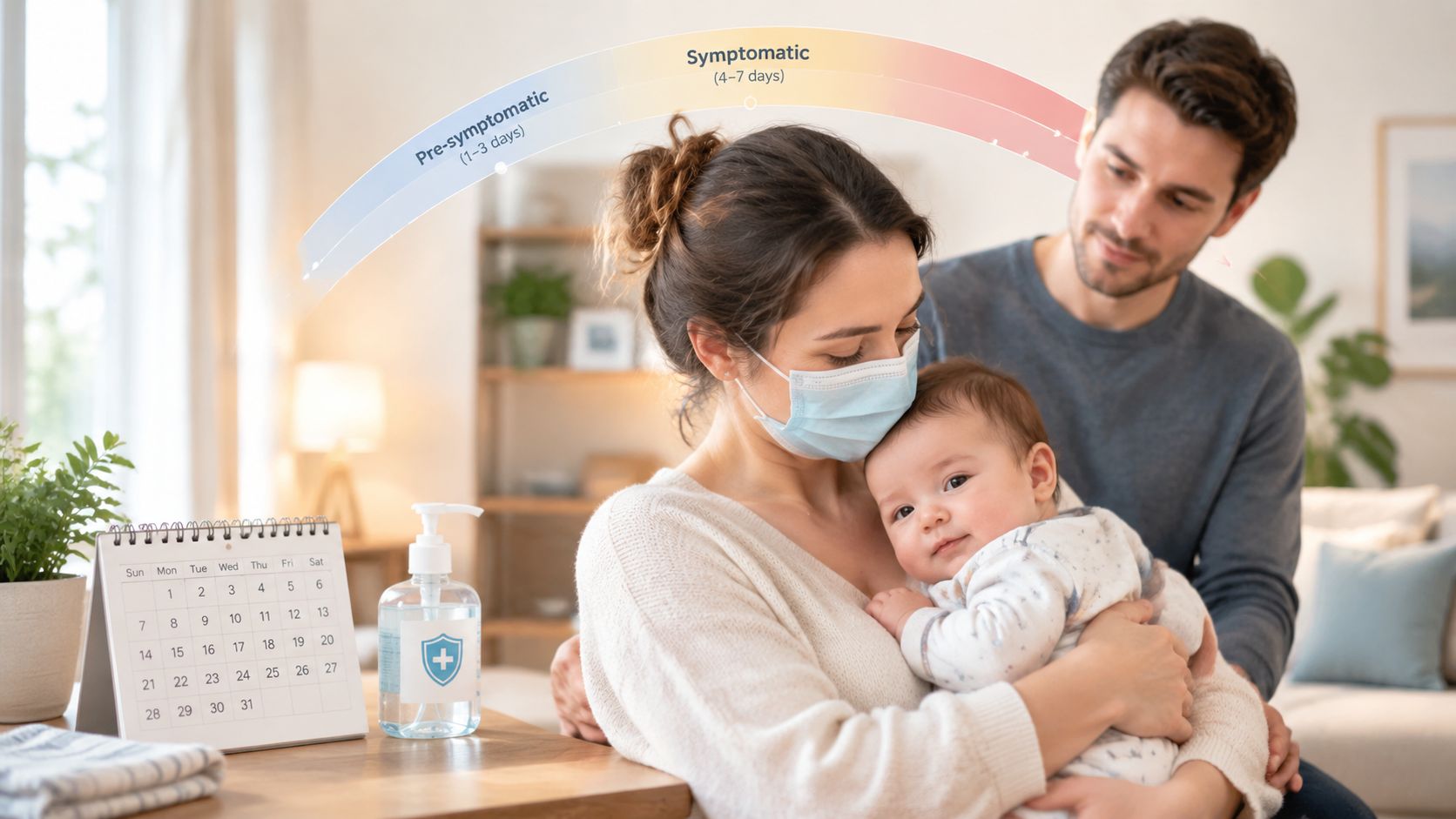
    People with hand, foot and mouth disease are usually most contagious during the first week, especially the first 5 days after symptoms begin. They can spread the virus a few days before symptoms appear and for weeks afterward through stool and respiratory shedding, even though the usual illness lasts 7 to 10 days.

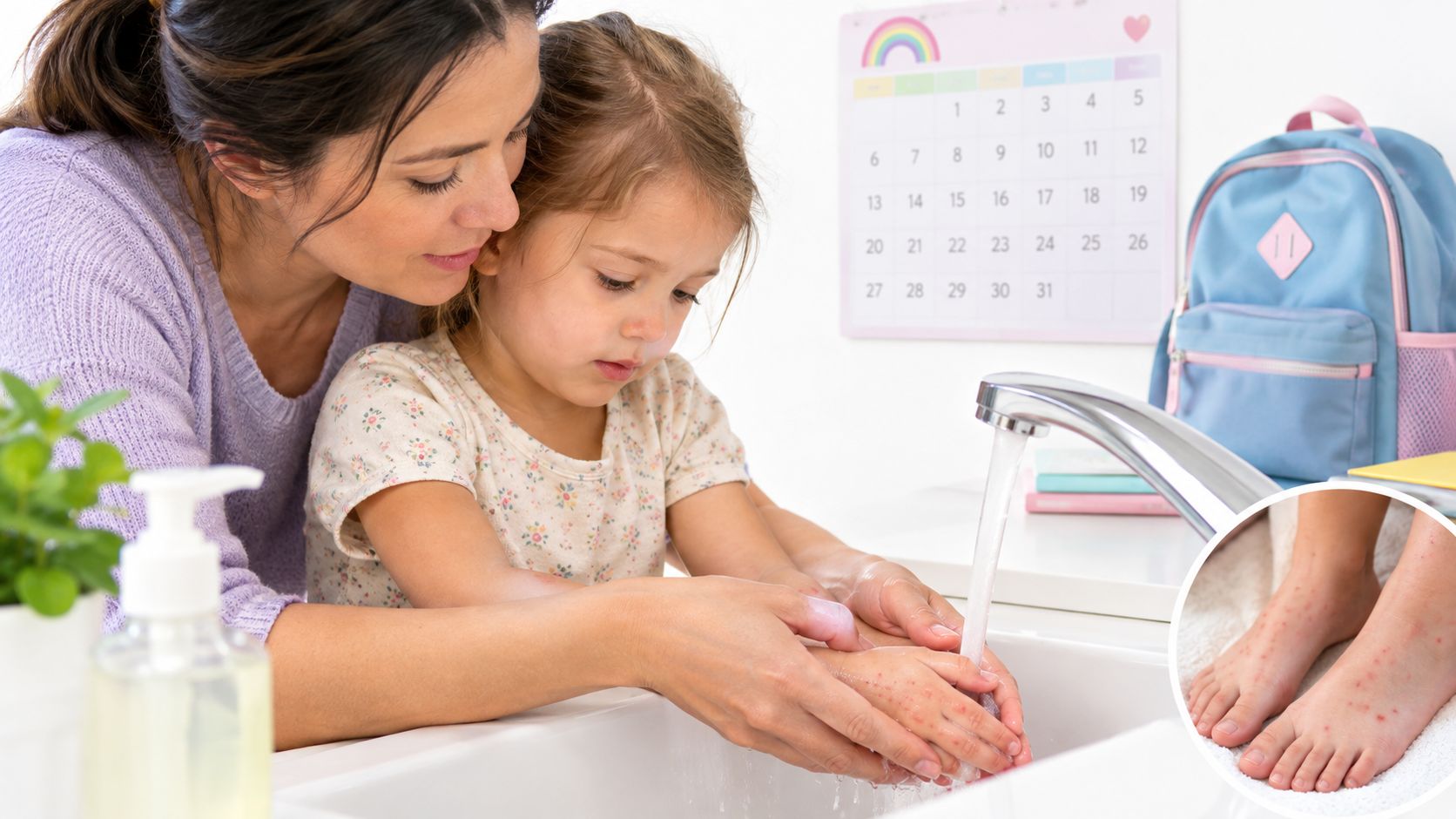
    A child may come home from daycare with a fever, sore mouth, and small blisters on the hands or feet. By the time the rash starts fading, parents often face a difficult question: is it safe to return to school, or could the child still infect classmates? Teachers and caregivers face the same uncertainty when a child seems well enough to participate but still needs help with toileting or diaper changes.

    The answer depends on two different timelines. Visible illness usually improves within the typical recovery period, while viral shedding can continue after the fever, mouth sores, and rash look better. Understanding that difference helps families make sensible return-to-school decisions without treating every lingering risk as a reason for indefinite isolation.

    Introduction to How Long Hand Foot Mouth Stays Contagious

    Consider a preschooler who develops a fever on Monday, mouth sores on Tuesday, and a rash on Wednesday. The child may be at especially high risk of spreading the virus during the early illness, when saliva, nose and throat secretions, blister fluid, and stool can carry it. By the following week, the child may be eating, playing, and sleeping normally, yet hygiene after toileting still matters because the virus can remain in stool well beyond visible recovery. The CDC's overview of hand, foot, and mouth disease describes this gap between symptom improvement and continued transmission.

    That gap explains why the question “how long is hand foot mouth contagious” doesn't have one simple answer. The highest-risk period comes early, but the full infectious window can extend beyond the rash. A child who feels better isn't automatically shedding nothing, and a child who still has a rash isn't necessarily in the same phase of illness as they were at the beginning.

    The simple timeline

    HFMD can spread before symptoms begin, is most likely to spread during the early days after symptoms start, and may continue to spread after recovery through respiratory secretions and stool. The NHS guidance summarized in public-health materials notes that people may start spreading the virus a few days before symptoms and are most likely to spread it during the first 5 days after symptoms begin.

    The rest of the timeline requires more nuance. Respiratory shedding may continue for 1 to 3 weeks, while stool shedding can persist for weeks or months. Those routes don't carry the same practical risk in every situation, but they explain why careful handwashing remains important after a child returns to normal activities.

    Practical rule: Recovery changes what a child can do, but it doesn't instantly erase every route of transmission.

    This distinction is useful for parents, teachers, childcare staff, and employers. It supports a balanced approach: keep someone home when they can't participate safely or has active symptoms that require exclusion, then continue strong hygiene after return.

    Understanding the Hand Foot Mouth Timeline From Exposure to Recovery

    HFMD begins after a person encounters an enterovirus and the virus has time to multiply before symptoms become noticeable. That quiet interval is the incubation period. It acts like a delayed message: exposure happens first, but the fever, sore throat, reduced appetite, mouth sores, and rash appear later. For a plain-language explanation of this stage, see what an incubation period means.

    The important point is that symptoms don't mark the beginning of infection. A person may already be able to spread the virus before anyone realizes they're ill. This is one reason a childcare outbreak can seem to appear suddenly, with several children becoming sick close together even though families followed normal routines before the first fever was recognized.

    A timeline graphic illustrating the stages of Hand Foot Mouth disease from initial exposure to full recovery.

    A fading echo rather than an on-off switch

    Early symptoms often include fever, sore throat, reduced appetite, or general discomfort. Mouth sores and skin changes then become more obvious, which makes the illness easier to identify. The visible phase usually improves within 7 to 10 days, as described by the CDC's HFMD information.

    Think of symptoms as the loudest part of an echo. The fever and rash are easy to notice, but the sound can continue after the original event has passed. In the same way, the disappearance of blisters doesn't prove that all virus has left the body.

    Four stages to keep in mind

    1. Exposure and incubation: The virus enters the body, but the person may look completely well.
    2. Early symptoms: Fever, throat discomfort, and reduced appetite can appear before the classic rash.
    3. Peak visible illness: Mouth sores and spots or blisters on the hands, feet, and sometimes other areas become prominent.
    4. Clinical recovery: Energy and appetite return, but shedding can continue through respiratory secretions or stool.

    This model prevents a common mistake. Parents often use the rash as a calendar marker, yet the contagious period doesn't begin with the rash and doesn't necessarily end when the rash fades. The most useful decisions combine symptom status, ability to participate, local exclusion rules, and hygiene support.

    When You Are Most Contagious and How Long Shedding Lasts

    A child can look well at breakfast, develop a fever that evening, and already have exposed classmates. HFMD transmission follows a curve rather than an on-off switch. Spread may begin before symptoms, rise around the start of illness, and continue after the skin clears.

    Public-health guidance indicates that people may spread HFMD a few days before symptoms begin. The highest likelihood of transmission is usually during the first 5 days after symptoms start, with the first week generally carrying the greatest risk. Transmission can still occur as symptoms improve. The CDC information on HFMD transmission identifies saliva, respiratory secretions, blister fluid, stool, and contaminated objects as routes of spread.

    HFMD contagious timeline at a glance

    Phase Typical Timing Contagious Risk
    Before symptoms A few days before symptoms begin Transmission can occur while the person looks well
    Early symptomatic illness First 5 days after symptoms begin Highest likelihood of spread
    First week Early illness through initial recovery Usually the most contagious phase
    Respiratory shedding 1 to 3 weeks Secretions may still carry virus
    Stool shedding Weeks to months Diapering and toileting remain transmission points

    The routes matter in different settings. Coughing, sneezing, saliva, and nose or throat secretions spread infection during close contact, including cuddling, kissing, shared meals, and classroom activities. Stool becomes a larger concern during diaper changes, toileting help, bathroom cleaning, and handling soiled clothing.

    A return to school can mean the child is well enough to participate, not that every route of transmission has ended.

    Why hand hygiene carries so much weight

    A caregiver can wash a child's hands before lunch and still miss the highest-risk moment: immediately after a diaper change or help in the bathroom. The CDC explanation of HFMD causes notes that enteric shedding can last longer, so handwashing after toileting and diaper changes remains an important control measure.

    Soap and water remove contamination from hands. Caregivers should also clean diapering areas, bathroom touchpoints, and shared objects according to their setting's procedures. The visible illness may have faded, yet routines involving stool still require care for weeks afterward.

    Why Some People Stay Contagious Longer Than Others

    A typical timeline helps with planning, but it can't predict the exact shedding pattern for every person. Virus type, the body's immune response, age, and underlying health can all affect how long viral material remains detectable or how easily someone can pass it on. That doesn't mean every child with HFMD needs a different exclusion rule. It means a blanket rule based only on the rash can miss important variation.

    Serotypes change the picture

    HFMD isn't caused by just one uniform virus. Different enteroviruses can produce similar symptoms, but their shedding patterns aren't identical. A meta-analysis found that positivity remained at roughly 50% around week 3 for CVA16 and CA6 cases and around week 4 for EV71 cases, with some stool shedding lasting more than 46 days. These findings are reported in the PubMed review of HFMD shedding.

    Those figures don't function as a personal countdown clock. A positive test or prolonged shedding doesn't automatically mean the person is equally infectious in every setting. Instead, the findings explain why researchers and public-health professionals avoid assuming that every serotype follows precisely the same course.

    Host factors also matter

    Young children often need hands-on help with eating, wiping noses, diapering, and toileting. That creates more opportunities for contact with secretions and stool than an older child or adult may have. Someone who is immunocompromised may also require individualized medical advice because the usual recovery pattern may not apply in the same way.

    A previous HFMD infection doesn't guarantee permanent protection. A 2025 reinfection-focused paper adds that people can get HFMD again, particularly when different serotypes such as EV-A71, CV-A16, or CV-A6 circulate. One episode can reduce uncertainty about the past illness, but it doesn't make future exposure harmless.

    A health infographic listing five safety criteria to follow before returning to school or work after illness.

    Use variation to guide caution, not fear

    Families shouldn't try to identify a serotype at home or extend isolation indefinitely without medical advice. A better response is to recognize the situations that increase contact risk:

    • Hands-on care: Diapering and toileting require meticulous handwashing.
    • Oozing lesions: Fluid from blisters can spread infection through direct contact.
    • Poor intake: Mouth pain may prevent safe participation and hydration.
    • Special health needs: Immunocompromised people may need personalized guidance.

    The safest timeline is not always the shortest one. It is the one that matches symptoms, activities, hygiene capacity, and professional advice.

    When Children and Adults Can Safely Return to School and Work

    Return decisions should focus on whether the person can participate safely, not on whether every trace of viral shedding has ended. Because respiratory and stool shedding can outlast symptoms, schools and workplaces generally use practical criteria such as fever resolution, improving sores, and the ability to manage normal activities. Families should follow the specific policy of the school, childcare program, employer, or local health authority.

    A child who still has significant mouth pain, can't drink comfortably, or needs more care than staff can provide should stay home. A child who is alert, able to take fluids, and ready to participate may be able to return when the applicable policy allows, while continuing careful hygiene.

    A return checklist

    • Fever has resolved: The child or adult should be able to function without relying on fever-reducing medication.
    • Sores are improving: Blisters should not be actively leaking, and exposed areas should be managed as appropriate.
    • Eating and drinking are possible: Severe mouth pain can make school or work unrealistic.
    • Normal participation is realistic: The person should have enough energy for the day.
    • Hygiene support is available: Handwashing after toileting, diapering, and nose wiping remains essential.
    • Policy has been confirmed: Ask the school, childcare provider, employer, or clinician what exclusion criteria apply.

    For teachers and childcare staff, the key question is practical: can this person use the bathroom, wash hands, eat, and interact without requiring care the setting can't safely provide? Parents should tell the school about the diagnosis or suspected illness so staff can reinforce cleaning and hand hygiene without singling out the child.

    Adults who work in childcare, healthcare, food service, or other close-contact settings may need to discuss return timing with their employer. Anyone with worsening symptoms, difficulty maintaining hydration, or an unusual course should contact a healthcare professional. Caregivers who work with children can also find paediatric first aid with Cura Academy to strengthen their response to common childhood health situations.

    For broader guidance about recognizing when illness requires time away from shared settings, consult when to stay home sick.

    An infographic showing the transmission routes and prevention methods for hand, foot, and mouth disease.

    How Hand Foot Mouth Spreads and Proven Ways to Stop It

    HFMD can spread before symptoms appear, during the early illness, and after visible recovery. Several routes matter, so one habit cannot block every exposure. Saliva, nose and throat secretions, blister fluid, stool, contaminated objects, and surfaces can all carry the virus. In a daycare, it may travel from a runny nose to a toy, from the toy to a hand, and from the hand to a cup or mouth.

    Match each route with an action

    Saliva and respiratory secretions spread through close interaction. Do not share cups, utensils, food, or items placed in the mouth. Teach children to cover coughs and sneezes, then clean their hands after wiping a nose. This route helps explain why respiratory shedding can still matter after a child feels well.

    Blister fluid creates a direct-contact risk. Children should not pick at blisters. Cover lesions when practical, provided the covering does not irritate the skin. Wash hands after touching dressings, clothing, or bedding that may have contacted fluid.

    Stool requires a consistent caregiver routine because shedding may continue after recovery. Wash hands with soap and water after every diaper change or toileting assist. Clean the changing surface before another child uses it.

    Objects and surfaces become part of the chain when several children handle them. Shared toys, diapering areas, eating utensils, drinking cups, tables, door handles, and toy bins deserve attention, especially where hand-to-mouth behavior is common.

    An infographic showing how hand, foot, and mouth disease spreads and how to prevent its transmission.

    The daycare and home connection

    A household may clean the sick child's bedroom while overlooking the shared bathroom, high chair, tablet, or favorite toys. A daycare may remove visible messes but miss the changing-table edge, cup rack, or toy bin touched by several children. Prevention improves when adults trace the route from source to hand to mouth instead of treating cleaning as one general task.

    Use an appropriate disinfectant according to its label and the surface instructions. Keep cleaning products away from children, and never mix them. Handwashing remains important because cleaning surfaces cannot remove contamination from caregivers' hands at every contact.

    For a clearer explanation of how viruses spread, consider how each contact creates another possible link in the chain. Breaking even one link, especially hand-to-mouth transfer, can reduce opportunities for HFMD transmission.

    Key Takeaways on Contagious Period and Next Steps for Prevention

    The answer to how long hand foot mouth is contagious has two parts. The first is the high-risk period: spread can begin before symptoms, with the greatest likelihood during the first week and especially the first 5 days after symptoms begin. The second is the extended shedding period, when respiratory secretions may remain relevant for 1 to 3 weeks and stool shedding can continue for weeks or months.

    Visible recovery still matters. A child who is fever-free, comfortable enough to eat and drink, energetic enough to participate, and able to follow the setting's hygiene expectations may be ready to return under local policy. Return doesn't mean zero transmission risk, so adults should continue careful hand hygiene and cleaning practices.

    Keep these points in mind:

    • Don't use the rash alone as the calendar: Symptoms and shedding follow different timelines.
    • Prioritize the early illness period: Close contact and shared items carry greater concern when symptoms are active.
    • Treat toileting as a long-tail issue: Handwashing after diaper changes and bathroom assistance remains important after recovery.
    • Respect individual variation: Serotypes and health conditions can change the shedding pattern.
    • Communicate clearly: Tell schools and childcare providers what symptoms are present and follow their exclusion rules.
    • Seek medical advice when needed: Difficulty drinking, worsening illness, or special health circumstances deserve professional guidance.

    Understanding the timeline should make decisions clearer, not more frightening. Families can support recovery while reducing spread by combining sensible exclusion with reliable handwashing, careful diapering and toileting routines, and regular cleaning of shared objects and surfaces.


    Review your household or classroom routine today. Identify the main hand-to-mouth and toileting contact points, clean the shared surfaces and objects according to product directions, and share this guide with the parents, teachers, and caregivers who help protect children during HFMD recovery. For more virus education and prevention guidance, visit VirusFAQ.com.

  • Evidence Based Medicine: A Practical Guide for 2026

    You're in a clinic waiting room, scrolling through headlines while waiting for your appointment. One story says the newest COVID booster is worth getting. Another says masks do little. A third presents an antiviral as a breakthrough, while someone in the comments insists that older treatments work just as well. Your clinician has limited time, and the studies behind those claims are more complicated than their headlines suggest.

    This is the everyday problem evidence based medicine is designed to address. It doesn't offer a permanent answer for every virus, vaccine, or treatment. Instead, it gives clinicians and patients a disciplined way to ask better questions, judge research, and adapt decisions when stronger or newer evidence appears. That skill matters whether you're evaluating a respiratory-virus recommendation, reading how epidemiological surveillance works, or deciding which claims deserve your attention.

    Why Evidence Based Medicine Matters Right Now

    Health decisions now arrive through several channels at once. A public-health agency may update guidance, a preprint may circulate widely before peer review, and a social-media post may reduce a complex trial to a confident sentence. The volume creates a practical problem: more information doesn't automatically produce better decisions.

    Evidence based medicine helps separate three questions that headlines often blend together:

    • Does an intervention work under study conditions?
    • How large and clinically meaningful is the benefit?
    • Does the finding apply to this person, community, or outbreak?

    Consider a patient asking whether a COVID booster is worthwhile. A careful answer depends on the patient's age, previous infections, immune status, circulating variants, vaccine availability, and tolerance for possible side effects. A single headline can't combine those factors responsibly. A clinician can use research evidence as a starting point, then interpret it in the patient's actual setting.

    Practical rule: A study can be scientifically credible and still be a poor fit for the person making the decision.

    The same reasoning applies beyond SARS-CoV-2. Influenza A, including H1N1 and H5N1, hepatitis viruses, norovirus, rotavirus, rhinoviruses, and herpesviruses create different questions about transmission, diagnosis, prevention, and treatment. Evidence based medicine doesn't make those viruses interchangeable. It helps you choose a study design and outcome that match the question.

    By the end of this guide, you should be able to turn a news claim into a focused question, identify what kind of evidence supports it, spot important limitations, and discuss the result more clearly with a clinician. You'll also have a repeatable way to respond when guidelines change, rather than treating every update as proof that medical knowledge has failed.

    What Evidence Based Medicine Really Means

    Evidence based medicine is the conscientious, explicit, and judicious use of current best evidence for decisions about individual patient care. It combines external research with clinical expertise, while accounting for the patient's values and circumstances, as described in this clinical reference on evidence based medicine.

    A GPS offers a useful analogy. The best current evidence resembles live traffic information. It tells you what routes appear open, where delays are likely, and how reliable the underlying information is. Clinical expertise resembles the driver's knowledge of local roads. A map may recommend a route, but an experienced driver knows about a difficult turn, a closed entrance, or a road that doesn't suit the vehicle.

    The patient's preferences provide the destination. Someone may prioritize avoiding hospitalization, minimizing side effects, reducing the chance of transmitting infection to a vulnerable relative, or avoiding an intervention that conflicts with personal values. A route can be technically efficient and still fail if it doesn't lead to the destination the traveler chose.

    The three parts of the decision

    These elements work together rather than competing with one another.

    • Research evidence asks what happened in properly conducted studies and how confident we should be.
    • Clinical expertise helps interpret whether the study population, dose, timing, and outcome resemble the patient in front of the clinician.
    • Patient values determine which benefits and harms matter most in the final choice.

    This approach differs from eminence-based medicine, where an authority's reputation may carry more weight than transparent evidence. Expertise still matters in EBM, but it doesn't receive a free pass. A respected clinician should be able to explain the evidence, its uncertainty, and the reasons a recommendation fits a particular patient.

    EBM is also a recalculation process. New vaccine formulations, viral variants, diagnostic tests, and antiviral trials can change the route. The goal isn't to preserve an old recommendation forever. The goal is to update the decision when the evidence, clinical context, or patient's priorities change.

    You can use this three-part lens whenever you read a study or guideline: What does the research show, how does it fit this situation, and what does the patient want to achieve?

    A Short History of Evidence Based Medicine

    Evidence based medicine developed through several connected efforts rather than one sudden invention. Earlier work in clinical epidemiology at McMaster University began in 1981, creating a foundation for teaching clinicians how to evaluate research instead of relying mainly on unsystematic experience. The label came later.

    A widely cited history traces the formal introduction of the term to a 1991 editorial in the ACP Journal Club, which argued for a more explicit shift from intuition and tradition toward the best available research evidence. This historical account of evidence based medicine describes the movement as a sequence: critical appraisal in the early 1980s, naming the approach in 1991, and building international systems for evidence synthesis afterward.

    The next major milestone came in 1993, when the Cochrane Collaboration was founded in Oxford. Its purpose was to prepare, maintain, and disseminate systematic reviews of randomized controlled trials. The collaboration began with a network spanning 13 countries, while an account of the first Cochrane Colloquium records 77 attendees from 11 countries at the meeting that helped establish its early international base. These figures are documented in this history of evidence based medicine and the Cochrane Collaboration.

    Cochrane's infrastructure gave clinicians something individual experts couldn't provide alone: coordinated summaries of large bodies of research. A handbook history records that the Cochrane Pregnancy and Childbirth Database had electronic publication in 1989 and developed into the broader Cochrane Database of Systematic Reviews in 1995, as described in the same historical reference.

    Why the timeline still matters

    The history explains why modern EBM emphasizes methods. First, clinicians learned to question unsupported authority. Then researchers developed ways to test interventions more fairly. Finally, international groups built systems to find and summarize those studies.

    That sequence now influences internal medicine, nursing, public health, guideline development, and virology. During fast-moving outbreaks, researchers and policy teams often have to make decisions before evidence becomes complete. The historical lesson is useful because it encourages neither blind confidence nor blanket distrust. It asks decision-makers to state what is known, what remains uncertain, and what new information could change the recommendation.

    Levels of Evidence and Study Designs Explained

    A study's design tells you what kind of question it can answer well. A clinician asking whether a virus causes a particular illness needs a different design from one asking whether an antiviral prevents hospitalization. Readers often call this arrangement an evidence pyramid, but the hierarchy is a guide, not a substitute for judgment.

    At the lower end, expert opinion can provide useful clinical context, especially when evidence is sparse. Its weakness is that memory, authority, and personal experience can distort judgment. Case reports and case series can flag unusual symptoms, unexpected adverse events, or a possible new pathogen, but they can't reliably establish how often an outcome occurs or whether a treatment caused it.

    Observational studies follow people without assigning the intervention. Cohort studies can compare vaccinated and unvaccinated groups, while case-control studies can compare people with an outcome to those without it. These designs can study real-world populations and questions that would be difficult or unethical to randomize, but confounding can make groups differ in important ways beyond the intervention itself.

    Comparing common designs

    Level Study Design Best Used For Main Weakness
    Foundational Expert opinion Context, clinical experience, urgent interpretation Authority and personal bias
    Early signal Case report or case series Unusual presentations or possible safety signals No reliable comparison group
    Real-world association Cohort or case-control study Risk factors, prognosis, effectiveness in practice Confounding and selection bias
    Strong intervention test Randomized controlled trial Comparing treatments or preventive measures Eligibility criteria may limit applicability
    Evidence synthesis Systematic review or meta-analysis Combining relevant studies and examining consistency Inherits weaknesses from included studies

    Randomized controlled trials improve comparability by assigning participants to intervention or comparison groups. Randomization helps balance known and unknown confounders, while blinding can reduce biased treatment or outcome assessment. Still, a trial may be too small, too short, or too selective to answer questions about rare outcomes, long-term effects, or people with complex illness.

    Systematic reviews use an explicit search and selection process to gather relevant studies. Meta-analysis can pool results, increasing statistical power and precision, reducing random error, and revealing heterogeneity, bias, and evidence gaps that an individual trial may miss, as explained in this review of meta-analysis in clinical research.

    The highest level of evidence isn't automatically the best answer. The best answer comes from the design that fits the question and has been conducted carefully.

    For teams handling complex research records, a voice-to-ELN for decision support may help capture observations and reasoning. It doesn't replace appraisal. It supports a transparent record of what was considered and why.

    The Five Step EBM Workflow in Practice

    Suppose an older adult asks whether a high-dose influenza vaccine is appropriate. A useful workflow turns that broad question into a decision that can be searched, tested, and revisited.

    1. Ask. Convert the scenario into PICO: the patient or population, intervention, comparison, and outcome. For example, ask whether an older adult receiving a high-dose influenza vaccine, compared with a standard formulation, experiences better protection against clinically important influenza outcomes.

    2. Acquire. Search efficiently in PubMed and the Trip Database. Use terms for the population, vaccine formulation, comparator, and outcome. Search results need screening, not automatic trust. A tool such as this researcher's guide to AI tools can help organize a literature search, but any summary still needs checking against the original paper.

    3. Appraise. Ask whether the study used a credible design, enrolled the right population, measured meaningful outcomes, and handled missing data or bias appropriately. CASP checklists can structure critical appraisal, while the GRADE approach helps judge certainty across the body of evidence.

    A diagram illustrating the five-step evidence-based medicine workflow from clinical question to improved patient care outcomes.

    1. Apply. Combine the findings with clinical expertise, the patient's preferences, vaccine access, local influenza activity, and relevant medical conditions. A statistically persuasive result may matter less if the study population differs substantially from the patient.

    2. Assess. Follow the outcome and review whether the decision achieved its purpose. Assessment can also examine the reasoning process itself. If the patient's health changes or new evidence appears, restart at the question stage.

    For a practical guide to moving from abstract to useful evidence, consult this explanation of how to read a scientific paper. The workflow is iterative because clinical knowledge and viral conditions change.

    Applying EBM to Virology and Public Health

    Virology makes uncertainty visible. A laboratory study may show that a mask filters particles under controlled conditions, while a community study asks whether people wear masks correctly, for how long, and alongside which other measures. Those are related questions, but they aren't identical.

    Masks and respiratory transmission

    Mask evidence can include mechanistic studies, observational comparisons, and randomized trials. An informed reader should ask whether the study examined source control, protection for the wearer, household transmission, workplace exposure, or community infection. The result also depends on adherence, fit, ventilation, the virus involved, and the period in which the study took place.

    A mixed evidence base doesn't justify choosing the most convenient conclusion. It calls for a more precise statement, such as: the intervention may reduce risk under some conditions, but the size of the benefit depends on exposure and implementation. Public-health teams can use epidemiological data analysis to interpret those changing conditions rather than treating one study as a universal answer.

    Influenza antivirals

    Neuraminidase inhibitors for seasonal influenza offer another lesson. Early enthusiasm can look different after researchers pool trials, examine outcomes carefully, and distinguish symptom duration from prevention of serious complications. A meta-analysis can narrow an apparently broad claim by showing which patients benefit, which outcomes improve, and where uncertainty remains.

    The practical question isn't “Do antivirals work?” It's “For which patient, at what point in illness, against which outcome, and with what trade-offs?” That wording prevents a laboratory mechanism or a persuasive anecdote from carrying more weight than the relevant clinical evidence.

    SARS-CoV-2 vaccines

    SARS-CoV-2 vaccine trials also show why recommendations must be updated. Trial results may establish benefits under particular conditions, but viral variants, prior immunity, circulating transmission, and available formulations can alter applicability. Guideline writers have to track new evidence while communicating what remains stable.

    The same framework applies to vaccine safety signals and to prevention decisions involving influenza A, H5N1, hepatitis B, or other viruses. Assess effect size, certainty, applicability, and equity together. An intervention that performs well in a controlled study may be harder to access, accept, or deliver in the community where it's most needed.

    Limitations and Honest Critiques of EBM

    A rigorous EBM process can still mislead. The method is only as trustworthy as the studies it finds, the outcomes researchers report, the assumptions clinicians make, and the fit between the evidence and the patient.

    Four ways evidence can fail

    Publication bias can hide disappointing findings. Researchers, sponsors, or journals may give less attention to negative or inconclusive antiviral and mask studies. Selective outcome reporting creates a related problem when a study measures many outcomes but emphasizes only the favorable result. A systematic review can't fully correct a gap if the missing studies never become visible.

    Funding and conflicts of interest can influence study design, interpretation, and presentation. The literature describes concerns about biased trial selection, manipulated design, and selective publication, particularly in industry-funded research, as discussed in this review of evidence based medicine's limitations. Financial disclosure doesn't prove that a result is wrong, but it tells readers to examine methods and reporting with care.

    Guideline lag creates another danger. Outbreaks and variants can change faster than committees can complete searches, consultations, and updates. Recommendations may therefore reflect the strongest evidence available at the time rather than the strongest evidence that will appear later.

    Average effects don't describe every patient. Trial participants may differ from someone with multiple conditions, altered immunity, pregnancy, or an unusual viral genotype. Reviews have long noted that randomized trials and meta-analyses often describe an average patient, while evidence can be weaker for diagnosis, prognosis, causes of illness, and preference-sensitive decisions, as explained in this review of applying evidence to individual patients.

    An infographic detailing four key limitations and potential pitfalls of evidence based medicine in clinical practice.

    Consider applying adult influenza antiviral findings to a severely immunocompromised child, or extending remdesivir evidence to a pregnant patient. The biological question may be related, but dosing, immune response, safety priorities, and outcome risks can differ. A clinician may need indirect evidence, pharmacology, specialist input, and careful monitoring.

    Recognizing these weaknesses isn't cynicism. It's mature EBM. The responsible response to uncertainty is to label it, seek better evidence, disclose assumptions, and avoid presenting a conditional finding as a universal rule.

    Trustworthy Resources and Your Next Steps

    Use the five-step sequence in one sentence: ask a focused question, search for evidence, appraise it critically, apply it with the patient, and assess the outcome.

    A practical checklist can keep the process manageable:

    • Frame the question: Use PICO to identify the population, intervention, comparison, and outcome.
    • Search systematically: Start with PubMed, PubMed Clinical Queries, or the Cochrane Library.
    • Filter by design: Match randomized trials, cohort studies, diagnostic studies, or reviews to the question.
    • Appraise transparently: Use CASP for critical appraisal and the GRADE handbook for certainty judgments.
    • Check reporting quality: Consult the EQUATOR Network when evaluating how researchers reported a study.
    • Verify public-health guidance: Compare recommendations from the CDC and WHO with the date, population, and virus addressed.
    • Discuss the decision: Bring the evidence to a clinician and explain which benefits, harms, and practical constraints matter to you.
    • Reflect afterward: Record what happened and whether new evidence changes the decision.

    Visual learners may also benefit from reputable critical-appraisal YouTube channels, provided the presenters identify their sources and distinguish evidence from opinion. For virus-specific background, VirusFAQ.com offers accessible and scientific articles about viral characteristics, transmission, identification methods, and prevention.

    A checklist infographic titled Trustworthy Resources and Your Next Steps outlining clinical evidence-based practice procedures.

    EBM improves with practice. Choose one recent headline about a mask, vaccine, antiviral, or outbreak measure this week, write its PICO question, find the original study, and note what would make the result more or less applicable to you. Then discuss your conclusion with a qualified clinician before changing treatment or prevention plans.


    Pick one viral-health claim you've recently seen, run it through the five-step workflow, and save the original study alongside your notes. If the decision affects vaccination, antiviral treatment, pregnancy, immune suppression, or a serious infection, contact a healthcare professional for individualized guidance rather than relying on a headline alone.

  • Contagious Period for RSV How Long You Spread It

    Individuals with RSV are contagious for 3 to 8 days and may become contagious 1 to 2 days before symptoms begin. Infants and immunocompromised people may remain contagious for 4 weeks or longer.

    A child can bring home what looks like an ordinary cough, play normally, and share toys with siblings before anyone suspects RSV. By the time the first runny nose appears, another family member may already have been exposed. That timing is why the contagious period for RSV matters for parents, childcare workers, clinicians, and anyone caring for an infant or a person with weakened immunity.

    RSV spreads through close contact and respiratory secretions, so households, classrooms, and childcare rooms give it many opportunities to move from person to person. The practical question isn't only, “How long does RSV last?” It's also, “When did contagiousness begin, and who might still be spreading it after feeling better?”

    Introduction to the RSV Contagious Window

    A preschooler returns home with mild congestion but no fever. The child hugs a parent, plays with a baby sibling, and goes to childcare the next morning. A day later, the parent starts coughing. Soon afterward, the infant develops feeding difficulty or noisy breathing. There may be no obvious moment when RSV entered the household, because transmission can begin before illness looks recognizable.

    For a typical infection, the contagious window lasts 3 to 8 days, and a person may spread RSV 1 to 2 days before symptoms appear. The CDC's RSV symptom guidance states that symptoms usually appear 4 to 6 days after infection. Some infants and immunocompromised people can continue spreading RSV for 4 weeks or longer, giving households a longer protection window to manage.

    Two timelines overlap. The visible timeline starts with signs such as coughing, a stuffy nose, or fever. The biological timeline can start while the infected person still feels well. This overlap explains why a family may identify the illness only after a baby, older adult, or immunocompromised relative has already been exposed.

    Why early spread causes confusion

    Presymptomatic transmission allows RSV to move before anyone recognizes illness. A child may still have enough energy for school, while an adult may dismiss a mild throat irritation. Once symptoms become clear, close contacts may already need monitoring and added protection.

    A known exposure or new respiratory symptoms should prompt practical changes. Avoid close contact with vulnerable people, improve hand hygiene, clean frequently touched surfaces, and consider keeping the symptomatic person apart from infants when possible. These steps cannot prevent every infection, but they can reduce opportunities for spread during the uncertain early window.

    The RSV overview from VirusFAQ.com offers broader background on the virus. For household decisions, the key question is more specific: could this person still spread RSV to a baby, older adult, or immunocompromised relative?

    Practical rule: Feeling well does not prove that a person is not contagious, especially after a known exposure or during the earliest phase of respiratory symptoms.

    How RSV Incubation and Contagiousness Fit Together

    A child can seem ready for school in the morning, then develop a cough that evening. RSV transmission may already have been possible during that quiet period. The timing makes more sense when infection is viewed as overlapping stages, not a switch that turns on with the first cough.

    Stage one begins with exposure

    Exposure happens when RSV reaches another person through close contact or contaminated respiratory material. Symptoms usually do not appear immediately. During incubation, the virus is developing while the exposed person may continue normal activities and interact with family members.

    RSV symptoms usually appear 4 to 6 days after infection, according to guidance on RSV symptoms. Incubation is the quiet interval between exposure and noticeable illness. It does not identify the exact start of contagiousness, because transmission may begin before symptoms are obvious.

    Stage two includes the hidden overlap

    A person may become contagious 1 to 2 days before symptoms begin. During this presymptomatic phase, someone might eat with the family, work, attend school, or care for a baby without realizing that RSV can spread.

    That is why “I wasn't sick yet” does not necessarily mean “I couldn't have spread RSV.” A household may only recognize the risk after the first cough, even though close contact has already occurred. Families protecting an infant or immunocompromised relative should treat a known exposure or new respiratory symptoms as a reason to reduce close contact and strengthen hand hygiene.

    Stage three is early symptomatic illness

    After symptoms appear, coughing, sneezing, nasal discharge, and face-to-face caregiving can create more opportunities for transmission. The usual contagious period is 3 to 8 days, with risk often greatest early in illness. Feeling better one morning does not by itself prove that spread has ended, particularly for infants or people with weakened immune systems.

    A timeline graphic illustrating the stages of RSV infection from initial exposure to full recovery.

    Respiratory droplets and secretions can carry RSV during ordinary household contact. The explanation of how droplet transmission works can help families understand why shared air, hands, and frequently touched surfaces matter during this overlap.

    How Long You Stay Contagious by Age and Immune Status

    A healthy adult may be nearing the end of RSV shedding while an infant in the same home is still passing the virus to others. Age, illness severity, and immune function shape the timeline, so one household rule cannot fit every person.

    The evidence summary in this review of RSV viral shedding describes adults as typically shedding for about 3 to 7 days. Infants may shed for up to 14 days in mild illness and around 3 weeks in severe cases. Some immunocompromised patients may shed for months, which makes individualized clinical guidance important.

    Population Typical Contagious Period Extended Shedding Notes
    Healthy adults About 3 to 7 days Symptoms may improve before shedding has ended.
    Older adults Variable, depending on health and immune status Age alone does not set one duration. Underlying conditions and recovery matter when protecting contacts.
    Infants Up to 14 days in mild illness Severe illness may be associated with shedding for around 3 weeks.
    Immunocompromised people May last 4 weeks or longer Some patients may shed for months, so clinicians may guide precautions and testing.

    Why infants need a different plan

    Infants need adults for feeding, bathing, carrying, and soothing. Those repeated close interactions make full separation difficult, even when the baby appears more alert or comfortable. A brighter mood is not a reliable sign that shedding has stopped, especially after severe illness.

    Protection therefore extends beyond the infant. Parents, siblings, childcare workers, and vulnerable relatives may all be exposed. During the longer infant window, households should combine hand hygiene, cleaning, ventilation, and distance from high-risk visitors instead of using the child's mood as a measure of infectiousness.

    Why immune status changes the timeline

    The immune system helps control and clear RSV. Weakened immune function can allow shedding to continue beyond the usual adult window. Months of shedding are possible for some immunocompromised patients, though they are not inevitable for everyone in this group.

    The practical question for clinicians is, “Who is infected, how severe was the illness, and who could be exposed?” A healthy adult recovering from a mild illness may need a shorter set of precautions than an immunocompromised patient, whose shedding requires a more individualized plan. Families should follow the treating clinician's advice when prolonged shedding is possible, particularly around newborns, older adults, or other people at higher risk.

    What Makes the Contagious Period Longer or Shorter

    A baby may seem brighter while still shedding RSV, while a healthy adult with a mild cold-like illness may be nearing the end of the usual window. Illness severity, immune function, age, and symptom trajectory help explain why people in one household can stop shedding at different times.

    The virus and the host shape this timeline together. Age and immune status matter because they affect how quickly the body controls and clears infection. That difference should guide household decisions about close contact, visitors, and care.

    Severity changes the practical risk

    Severe bronchiolitis in an infant calls for greater caution. An RSV shedding review reports that infants may shed for up to 14 days in mild illness and around 3 weeks in severe illness (evidence summary). A child with more severe disease may therefore need protection around newborns, older adults, and immunocompromised relatives for longer than a healthy adult with mild symptoms.

    Symptoms change the number of opportunities for spread. Heavy coughing and frequent nasal secretions can carry respiratory material onto hands, clothing, toys, and nearby surfaces. As those symptoms ease, exposure opportunities may decrease, but improvement does not prove that shedding has ended.

    For families, the child's energy level is only one clue. The illness pattern, age, and health of people nearby matter too.

    Fever and improvement are useful, but incomplete

    The CDC advises staying home while respiratory symptoms are not improving and until the person has been fever-free for at least 24 hours. It then recommends extra precautions for the next 5 days, since transmission may still occur (CDC causes and prevention guidance). This is a practical public-health rule, not laboratory proof that all virus has disappeared.

    RSV is generally most contagious during the first week or so after infection, according to Mayo Clinic information. That makes the early period the time for the strongest household precautions, while leaving room for a later tail of shedding.

    A visual guide outlining health precautions like staying home, hygiene, and masking during a contagious period.

    Improvement shows that the illness is heading in a better direction. It does not guarantee that every transmission risk has ended.

    Around a newborn or immunocompromised household member, keep precautions stronger for longer and ask the treating clinician how the person's condition changes the plan. Symptoms may be enough for routine decisions in a healthy adult, but prolonged shedding may require individualized guidance.

    Isolation and Infection Control While You Are Contagious

    A child becomes ill overnight, while a parent still needs to provide meals, comfort, and bathing. In that situation, infection control means reducing close exposure wherever possible, not attempting perfect separation. Combining several small barriers works better than relying on one action.

    Start with the stay-home rule

    Stay home while symptoms are worsening or not improving. Follow CDC guidance to resume normal activities when symptoms are improving and the person has been fever-free for at least 24 hours, then use added precautions for the next 5 days. This public-health timetable helps guide daily decisions, but it does not prove that all RSV transmission risk has ended.

    School, daycare, and workplace policies may add their own requirements. An adult who can avoid close contact may return under a different plan from an infant who needs frequent hands-on care. Infants and immunocompromised people may shed RSV longer, so households should keep protective measures in place for longer and ask the treating clinician for individualized advice.

    Reduce exposure inside the home

    Wash hands after wiping a nose, handling tissues, feeding a child, or touching shared objects. Use separate towels when practical, improve airflow, avoid face-to-face contact, and postpone visits from vulnerable people while symptoms continue.

    Clean frequently touched hard surfaces, including door handles, light switches, faucet handles, changing areas, and shared toys. Use a suitable disinfectant according to its label. Never mix cleaning products.

    • Mask around vulnerable people: Wear a well-fitting mask when close contact cannot be avoided, especially near infants, older adults, or immunocompromised people.
    • Protect caregiving routines: Have one healthy caregiver manage feeding and soothing when possible, while another adult handles shared household tasks.
    • Keep supplies separate: Do not share tissues, cups, towels, or personal items during active illness.
    • Ask before returning: A clinician can help interpret prolonged symptoms or suspected extended shedding in an infant or immunocompromised patient.

    The practical goal is fewer close exposures, for less time, with less face-to-face contact. That approach protects vulnerable household members even when complete isolation is impossible.

    Testing Timing and When to Retest for RSV

    A parent tests a child the morning after a close exposure and gets a negative result. That result may be too early to settle the question. Symptoms often appear 4 to 6 days after infection, so testing immediately after exposure can occur before enough virus is present or before illness begins.

    Match testing to the timeline

    After a known exposure, watch for respiratory symptoms during the following days. Once symptoms start, ask a healthcare professional whether testing is appropriate, which test fits the situation, and how the person's age and immune status affect contact with vulnerable people.

    A negative result is one piece of evidence, not a clearance pass. Collection quality, test type, and the amount of virus present can all affect the result. See this comparison of antigen tests versus PCR to understand why different methods may produce different answers.

    Retesting may make sense when symptoms develop after an early negative result, symptoms continue, or a clinician believes the first sample did not match the illness timeline. The decision should follow the person's symptoms, exposure risk, and likelihood of prolonged shedding, especially for infants and immunocompromised people.

    When a clinician should guide the plan

    Seek prompt professional advice for an infant with breathing difficulty, poor feeding, unusual sleepiness, or other concerning changes. Older adults and immunocompromised patients may also need an individualized plan because they can remain infectious longer than healthy adults.

    If reaching a clinic is difficult, at-home blood draws may help with some diagnostic or monitoring needs. A blood draw does not replace a respiratory swab when RSV testing requires a respiratory sample, so ask the provider which specimen and timing are appropriate.

    Do not use one negative test as permission for a recovering child to have close contact with a high-risk relative. Interpret the result alongside symptoms, timing, age, immune status, and the household's exposure risk.

    Prevention Tips That Reduce RSV Spread at Home and Beyond

    RSV prevention works best as a layered routine. Handwashing removes virus from hands before they reach the eyes, nose, or mouth. Avoiding close contact during active illness lowers direct exposure, while ventilation and masking can reduce risk when separation isn't practical.

    Clean frequently touched hard surfaces as part of the same plan. Use EPA-registered disinfecting wipes according to the product label, including directions about contact time and compatible surfaces. Wipes can be convenient for doorknobs, faucet handles, changing tables, and other high-touch areas, but cleaning shouldn't replace hand hygiene or staying home when sick.

    A household routine that people can follow

    • Wash hands at key moments: Clean hands after nose wiping, coughing, diaper changes, and contact with shared surfaces.
    • Limit close contact: Avoid kissing, face-to-face play, and unnecessary visits while someone is symptomatic.
    • Protect high-risk people: Keep infants, older adults, and immunocompromised contacts away from active illness whenever possible.
    • Clean shared objects: Wipe hard, frequently touched toys and surfaces, following label instructions.
    • Communicate early: Tell household members and caregivers when symptoms begin so they can make informed contact decisions.
    • Watch the recovery: Feeling better is encouraging, but longer precautions may be appropriate for infants and people with weakened immunity.

    The central lesson about the contagious period for RSV is simple but easy to miss: transmission can begin before symptoms, and shedding doesn't last the same amount of time for everyone. Healthy adults may follow a shorter course, while infants and immunocompromised people can carry a longer transmission tail.

    For continuing virus education, visit VirusFAQ.com and subscribe to updates. At home, add labeled disinfecting wipes to your infection-control supplies, use them correctly on appropriate hard surfaces, and combine them with handwashing, ventilation, masking, and sensible distance from vulnerable people.


    If someone in your household has RSV symptoms, identify the vulnerable contacts first, separate the sick person where practical, and contact a healthcare professional promptly for an infant, older adult, or immunocompromised patient. Use the timeline above to guide precautions today, and keep EPA-registered disinfecting wipes available for the high-touch surfaces your family shares most often.

  • Viral Cytopathic Effects: A Guide to Cell Culture Changes

    You've looked through a microscope at a cultured cell layer and noticed that the tidy sheet of cells no longer looks tidy. Some cells have rounded up, others have pulled loose, and a few may have fused into strange, oversized structures. That visual change is called a viral cytopathic effect, or CPE.

    CPE gives virologists a fast way to recognize that something has happened inside a cell culture. It can support virus isolation, help quantify infectious virus, and guide further testing. But it has a major limitation: a damaged-looking cell isn't a direct meter of viral replication. The appearance may reflect viral proteins, altered membranes, programmed cell death, or the host cell's attempt to stop the infection.

    What Viral Cytopathic Effects Really Mean

    On the third day after infection, a researcher tilts a flask beneath an inverted microscope. The uninfected control monolayer resembles a neat cobblestone street, with cells pressed closely together and attached to the plastic. The infected flask tells a different story. Cells have become round and bright, gaps have opened in the sheet, and some areas have detached completely.

    That visible departure from healthy cell structure and behavior is a cytopathic effect. The term covers structural changes caused by viral infection, including cell rounding, detachment, fusion, inclusion bodies, and lysis. The National Center for Biotechnology Information describes CPE as the set of structural changes produced in host cells by viral infection, while a virus that produces those changes is called cytopathogenic (this overview of viral cytopathic effects).

    The distinction matters:

    • CPE is the observable phenotype, or what the cells look and do.
    • Cytopathogenicity is the virus's capacity to produce that phenotype.
    • Viral yield is the amount of infectious virus produced.

    Those three ideas can overlap, but they aren't interchangeable. A culture can show dramatic CPE without producing proportionally more infectious particles, while another culture can contain replicating virus with little obvious morphological change.

    The historical turning point

    CPE became a formal virology milestone in 1949, when John F. Enders, Thomas H. Weller, and Frederick C. Robbins reported cytopathic effects in tissue culture. Their work helped establish in vitro virus cultivation as a core diagnostic method in modern virology (the historical and biological review of CPE).

    Before cell culture became central, researchers often had to rely on animal inoculation or wait for symptoms in an infected host. A reproducible change in cultured cells offered a new window into virus and cell interactions. Researchers could watch infection unfold in a controlled setting, compare virus preparations, and develop methods for isolation and quantification.

    Modern virology has also widened the meaning of CPE. It isn't limited to simple cell death. Visible changes may reflect necrosis, apoptosis, or host-defense-driven self-destruction pathways. In practice, the same infected monolayer can display several signs at once, so the microscope provides an important starting signal rather than a complete explanation.

    Common CPE Morphologies in Cell Culture

    A culture dish may look normal at first. Then a few cells become round, bright, and separated from the surface. Over the next observations, that small change may spread across the monolayer. Reading CPE means treating each visible pattern as a clue about stressed cell systems, not as a diagnosis by itself.

    A diagram illustrating common cytopathic effect (CPE) morphologies in cell culture including rounding, shrinkage, detachment, and vacuolization.

    Rounding and shrinkage

    Healthy adherent cells spread across the growth surface. During infection-related stress, they may retract their cytoskeleton and become round, smaller, and more refractile. The change resembles balloons losing their broad shape and pulling away from nearby balloons.

    The cytoskeleton supports cell shape, while adhesion structures keep the cell attached to the extracellular surface. Viral disruption of signaling, protein production, or adhesion can remove that spread-out architecture. Rounding may come before detachment, but its timing depends on the virus, cell line, inoculum, and observation conditions.

    Swelling and vacuolization

    Some infected cells enlarge or develop clear spaces called vacuoles. Disturbed ion channels and membrane permeability can alter the balance of water inside the cell, giving it a bloated, waterlogged appearance.

    The membrane normally controls movement into and out of the cell. Mechanistic work describes increased membrane permeability, osmotic swelling, and membrane alterations as processes that can visibly distort infected cells (the mechanistic review of CPE pathways). A vacuolated cell therefore signals altered cell physiology, but does not identify the virus or prove how much infectious virus the culture contains.

    Fusion and syncytia formation

    A syncytium is a large cell containing multiple nuclei because neighboring cells have fused. Under the microscope, it resembles soap bubbles joining across the surface of water.

    Viral or virus-modified membrane glycoproteins can appear at the cell surface and drive adjacent plasma membranes to merge. Syncytia are associated with paramyxoviruses and coronaviruses, whereas other virus families more often produce rounding or detachment (educational material on CPE patterns and infectivity measurement).

    Focal degeneration and lysis

    CPE may begin in localized patches. Infected cells deteriorate, nearby cells become involved, and the affected region expands into a focus or plaque-like area. Complete loss of membrane integrity can cause lysis and detachment of the monolayer.

    These patterns show where damage is occurring and how it moves through the culture. They do not directly measure replication efficiency. Host-cell death pathways can intensify or reshape the visual result, so microscopy should be paired with a replication or infectivity assay when the question requires more than morphology.

    Microscope rule: Describe the morphology first, then test the mechanism. “Rounded and detached” is an observation. It is not yet an explanation.

    How Viruses Damage Cells at the Molecular Level

    The microscope shows the outcome of a molecular contest. Viral proteins redirect the cell toward virus production, while the cell activates defenses that can slow replication, shut down protein synthesis, or trigger self-destruction.

    Direct injury and the loss of cellular production

    Some viral structural or accessory proteins interfere directly with membranes, organelles, and intracellular transport. Viral protein accumulation can crowd the cytoplasm or nucleus and form visible inclusions. Other viral products change ion movement, damage membrane integrity, or disrupt the cytoskeleton.

    Viruses also need the host cell's manufacturing systems. When infection suppresses host macromolecular synthesis, the cell loses the ability to maintain its normal structure. A familiar example is poliovirus, whose 2A protease cleaves eIF4G, a host translation factor. The result is a shift away from normal cellular protein production and toward viral priorities.

    Apoptosis as a defensive sacrifice

    A cell may also choose to die. Sensors such as PKR detect infection-related signals and can contribute to stress responses and apoptosis. Programmed cell death removes the cellular environment that viruses need, limiting the opportunity for new particles to form and spread.

    That response creates a key interpretive problem. A severe CPE pattern may reflect an effective host defense rather than unusually efficient viral replication. Virology literature describes many CPE phenotypes as products of competing host-versus-virus pathways, including necrotic and apoptotic death (the review of virus-modulated cell death and CPE).

    Immune-mediated injury

    In a tissue or clinical setting, infected cells may also be damaged by cytotoxic T cells, cytokines, and other immune responses. The culture dish usually lacks the full immune environment, but host-response pathways can still influence what cells look like and how quickly they deteriorate.

    Syncytia illustrate why morphology can mislead. A virus may create syncytia because its glycoproteins have reached the plasma membrane and triggered fusion. That visible event depends on membrane behavior, not necessarily on the amount of infectious virus released.

    For a broader explanation of how viral mechanisms translate into disease, see how viruses cause disease.

    CPE Patterns Across Major Virus Families

    Different viruses leave different visual signatures, but the signature belongs to the virus-cell pairing, not to the virus alone. Cell type, culture conditions, inoculum, and timing all shape the result.

    Influenza A in MDCK cells may produce rounded, refractile foci that merge into larger patches. Hemagglutinin binding can alter cell interactions and contribute to clumping. Herpes simplex virus in Vero or HFF cells commonly produces rapid rounding and ballooning degeneration, with Cowdry type A intranuclear inclusions visible after H&E or Papanicolaou staining.

    HIV provides a different visual lesson. In primary lymphocytes and macrophage cultures, gp120-mediated membrane fusion can generate multinucleated giant cells. The syncytium is therefore a direct clue to envelope glycoprotein activity at the cell surface.

    Rotavirus in MA-104 cells may detach the monolayer without producing classical lysis. The mechanism involves NSP4-mediated calcium dysregulation, which changes intracellular signaling and cell integrity. Rhinovirus in H1-HeLa cells can produce shrunken, refractile, pyknotic cells, often clustered near the edge of the culture medium.

    Virus family Cell system Time-to-CPE Dominant morphology
    Influenza A MDCK Depends on culture conditions and assay timing Rounded, refractile foci and patch formation
    Herpes simplex virus Vero or HFF Depends on isolate and culture conditions Rounding, ballooning degeneration, intranuclear inclusions
    HIV Primary lymphocytes and macrophages Depends on infection and host-cell conditions Syncytia and multinucleated giant cells
    Rotavirus MA-104 Depends on inoculum and assay timing Monolayer detachment, often without classical lysis
    Rhinovirus H1-HeLa Depends on isolate and culture conditions Shrunken, refractile, pyknotic cells

    Reading the table correctly

    The table works as a prototype guide, not as a species-level identification tool. A herpesvirus-like pattern can support suspicion, but confirmatory testing must establish what caused it. Likewise, the absence of an expected pattern doesn't automatically exclude infection, particularly when the cell system isn't well suited to the virus.

    Measuring CPE to Quantify Infectious Virus

    A single flask can show that infection occurred. To estimate how much infectious virus is present, researchers compare many replicate cultures across controlled dilutions. The classic format is the endpoint dilution assay.

    A viral stock is diluted serially and added to replicate wells containing susceptible cells. At a defined observation point, each well receives a positive or negative CPE score. Researchers then estimate the dilution that infects 50% of cells or test systems, the TCID50 endpoint, using the laboratory explanation of TCID50, PFU, and CPE scoring.

    The 50% benchmark does not require every infected cell to die or every well to display identical damage. Spearman-Kärber and Reed-Muench interpolation methods estimate the endpoint from the pattern of positive and negative wells.

    TCID50 and PFU answer related questions

    A plaque assay places infected cells beneath a semisolid overlay, restricting the spread of progeny virions. Each localized infectious event can form a visible focus, which researchers count after staining with agents such as neutral red or crystal violet. The resulting plaque-forming units, or PFU, measure infectious virus through counted plaques.

    These readouts describe related but different outcomes. TCID50 uses the distribution of CPE across replicate units. PFU uses discrete, countable infection foci. Both depend on a susceptible cell system, consistent culture conditions, and a defined scoring window.

    A diagnostic flowchart showing the process of identifying viral cytopathic effects from a clinical specimen through culture methods.

    Practical caution: A visual endpoint depends on timing, multiplicity of infection, cell health, and operator judgment. Keep these variables consistent before comparing results.

    CPE-based quantification remains useful in vaccine research, antiviral screening, and serum neutralization tests. It turns a visible cell response into a reproducible statistical endpoint, while leaving room for interpretation because visible damage does not always map perfectly to viral replication. For added context, review viral culture methods.

    CPE in Modern Diagnostics and Laboratory Workflows

    Modern laboratories rarely treat CPE as a standalone answer. Instead, microscopy acts as an early triage signal inside a workflow that may include culture, staining, nucleic-acid testing, and sequencing.

    In shell vial culture, centrifugation can help bring specimens into closer contact with cells, after which virus-specific monoclonal antibodies may detect infection before overt CPE appears. Conventional tube culture remains useful when the virus grows more slowly or when the laboratory needs extended observation. These approaches can support detection of CMV, HSV, VZV, and respiratory viruses, but the readout often depends on immunostaining rather than morphology alone.

    Some agents require other signals. Hemadsorption can reveal viral effects on red-cell binding at the infected cell surface. Hemagglutination can detect the ability of released virus to agglutinate red blood cells. Interference assays can also help identify agents that don't produce clear CPE, including rubella and some influenza strains.

    Confirmation makes the result defensible

    When the culture appearance is ambiguous, laboratories can select a confirmation layer suited to the question:

    • Immunofluorescence panels detect viral antigens in cells.
    • Real-time RT-PCR detects viral nucleic acid with high analytical sensitivity.
    • Electron microscopy can reveal particle morphology in selected investigations.
    • Next-generation sequencing can characterize viral genetic material when targeted tests don't provide enough information.

    A diagram illustrating the six-step diagnostic workflow for detecting Carbapenemase-producing Enterobacteriaceae (CPE) in a laboratory setting.

    A clean culture doesn't end the investigation when infection remains plausible. The laboratory may extend incubation, use a different host cell, perform blind passage, co-cultivate the specimen with an indicator line, or move directly to molecular detection.

    These decisions sit alongside broader safety practices. Anyone handling potentially infectious specimens should also review practical clinical blood safety procedures, especially when work involves bloodborne pathogens and exposure prevention.

    CPE remains valuable because it tells staff where to look next. Guidance on combining culture with confirmatory methods is available in laboratory diagnosis of viral infections.

    Why Absence of CPE Does Not Mean Absence of Infection

    A clear-looking monolayer can still contain virus. “No CPE observed” means only that the chosen culture system did not show visible damage under the conditions and observation schedule used. It doesn't prove that no virus is present.

    Some viruses replicate or persist without strongly changing cell shape. HBV may be difficult to assess through conventional morphology, and some coronaviruses may show limited visible effects in low-passage cells. A restricted host range can prevent productive infection altogether in the selected cell line, while a low multiplicity of infection may leave too few cells visibly affected to trigger a confident microscopic call.

    Sample quality can complicate interpretation too. Toxic material in a clinical specimen may injure cells and obscure virus-specific patterns. Conversely, subtle cytoskeletal disruption, delayed syncytium formation, or apoptosis that develops later may be missed if the culture is examined only at an early timepoint.

    A responsible negative result needs context

    The appropriate follow-up depends on the suspected virus and the purpose of the test. Useful options can include:

    • Immunostaining, to look for viral antigen inside apparently healthy cells.
    • Hemadsorption, when infected cells may bind red blood cells without obvious CPE.
    • RT-PCR, to detect viral RNA when morphology is uninformative.
    • Electron microscopy, when particle visualization can answer a specific question.
    • Extended culture or blind passage, when the virus may need more time or a second round of amplification.

    The central distinction is simple: absence of visible CPE is not absence of infection. Careful diagnosticians treat morphology as one piece of evidence, weigh the cell line and assay design, and confirm a negative interpretation with an appropriate independent method when clinical or experimental suspicion remains.


    If you want clear, evidence-based explanations of viruses, transmission, culture findings, and practical prevention, explore more guides at VirusFAQ.com, and use the site's prevention resources to choose appropriate disinfecting wipes for routine surface hygiene.

  • Norovirus Incubation Period: Key Facts to Know

    The norovirus incubation period is typically 12 to 48 hours, most often around 24 to 36 hours. A person can feel perfectly well after exposure, then develop sudden vomiting or diarrhea within the next day or two.

    That timing creates a familiar household puzzle. One person becomes ill overnight, while someone else who shared the meal remains well. The difference isn't always the food itself. Exposure dose, individual susceptibility, viral strain, and the timing of contact can all change what happens next.

    The clock matters beyond curiosity. Families use it to decide whom to monitor. Schools and care facilities use it to watch for additional cases. Food services and healthcare teams use it to determine when exclusion, cleaning, and surveillance should continue. Understanding the incubation period helps you make safer decisions before symptoms appear and after they stop.

    The Hour That Changes Everything

    On a Sunday evening, a family eats together. The father feels normal through dinner, chats with everyone, and helps store the leftovers. By midnight, he is vomiting. His daughter ate the same dishes but wakes the next morning feeling fine.

    That difference can mislead the family. The exposure might have come from the meal, an earlier contact, or a contaminated surface. Her lack of symptoms is reassuring only for the present. It does not show that she avoided infection or that no further case will appear. The practical questions are when exposure occurred and how long to watch for symptoms.

    Norovirus can spread through a household quickly because only a small amount of virus may cause infection, as the CDC explains. A vomiting episode in a shared kitchen, bathroom, dining area, or care setting can contaminate several places and create repeated opportunities for contact.

    The timing problem appears in many settings:

    • Households: One person becomes ill while relatives wait to learn whether they were exposed.
    • Schools: A child leaves after vomiting, but classmates and staff may already have encountered the virus.
    • Restaurants: A food worker can feel well during a shift and become sick later.
    • Care facilities: Residents and caregivers may need monitoring after the first patient improves.

    Practical rule: Feeling well now describes the current moment. It does not prove that an earlier exposure caused no infection.

    For outbreak control, symptom timing guides action. If a person is exposed today, the household or facility watches through the usual incubation window. If a new case appears, that case can restart attention to later exposures. In practice, monitoring for two incubation periods after the last suspected exposure gives people time to detect cases that develop later, rather than ending precautions as soon as the first patient feels better.

    The incubation period is a framework, not a perfect prediction. It cannot identify the exact source of every illness or guarantee that every exposed person will become sick. The useful question is not “Who feels sick?” Ask instead: “When might exposure have occurred, and when can monitoring safely stop?”

    What the Incubation Period Actually Means

    A person can share a meal at noon, feel completely normal through the afternoon, then wake with nausea or vomiting overnight. That quiet interval is the incubation period, the time between catching a virus and developing the first symptom. Norovirus may already be multiplying while the person eats, works, attends school, or cares for someone without knowing an infection has begun.

    For norovirus, the usual window is 12 to 48 hours, according to CDC clinical guidance on norovirus. Outbreak research places the center of that range more precisely. An analysis of 1,022 outbreaks found a mean incubation period of 32.8 hours and a median of 33.5 hours. A systematic review of 2,540 observations estimated a median of 1.2 days for norovirus genogroups I and II, as reported in the peer-reviewed outbreak analysis.

    These figures describe a pattern, not a fixed appointment. Some people become ill sooner, some later, and some develop no noticeable symptoms.

    What the clock starts and stops

    The clock begins at exposure, not necessarily at the last meal you remember. Exposure can occur through contaminated food or surfaces, direct contact with an infected person, or particles released during vomiting. The clock ends when the first symptom appears, often nausea, vomiting, diarrhea, or abdominal discomfort.

    It does not measure the time until fatigue, appetite loss, or full recovery. Those belong to the illness and recovery stages, which follow the incubation period.

    For a broader explanation, see what an incubation period means.

    A six-step infographic explaining the incubation period of pathogens from initial exposure to the appearance of symptoms.

    This timeline supports practical outbreak decisions. A person who feels well has not necessarily passed the risk window, so households and facilities monitor after a suspected exposure rather than stopping precautions when the first patient improves. Watching through two incubation periods after the last suspected exposure allows later cases to appear before monitoring ends. Timing, not just feeling better, helps show when the group is in the clear.

    The range cannot identify every source or predict every case. It frames the useful question: when could exposure have occurred, and when can monitoring safely stop?

    Why the Clock Varies From Person to Person

    Two people can share a meal yet develop symptoms at different times. The difference does not automatically point to different illnesses. Incubation depends on the exposure each person received and on how each body responds.

    Dose changes the starting conditions

    The amount of virus entering the body can vary widely. A contaminated forkful of food may deliver more virus than touching a contaminated handrail and then touching the mouth. Norovirus has an estimated infectious dose of only 10 to 100 virions, a point described in the CDC prevention guidance.

    A larger inoculum may bring symptoms earlier, but particle count cannot produce an exact personal forecast. People almost never know how much virus they encountered. Dose is therefore one reason timelines differ, not a countdown calculator.

    Host biology adds another layer

    Age, previous exposure, immune response, and other biological features can influence whether infection becomes noticeable and how soon symptoms appear. Two people with similar exposure may have very different experiences. One may develop intense gastrointestinal symptoms, while another has a milder illness or no obvious symptoms.

    Blood group and prior immunity have been studied as possible influences, but neither provides a dependable household prediction. Someone should not assume they are protected because a family member became sick while they did not.

    Strains don't behave identically

    Norovirus includes multiple genogroups and strains. Genogroup II viruses, including recombinant variants associated with widespread outbreaks, can behave differently from genogroup I viruses. That variation is why outbreak investigators compare timing and symptoms across many cases instead of treating one person's incubation period as a universal rule.

    Factor Direction of effect Typical range change
    Exposure dose A greater dose may move symptoms earlier The window can shift toward the early part of the usual range
    Host factors Susceptibility and immune response may move symptoms earlier, later, or make them less noticeable No dependable individual adjustment
    Viral strain Different genogroups and variants may produce different outbreak patterns No fixed adjustment for a single person

    The 12 to 48 hour range remains more useful than a promise about one exact hour. It helps households and facilities decide how long to monitor after a suspected exposure, while recognizing that no single person's timing can mark the end of risk for everyone.

    Symptoms that have not started yet do not, by themselves, settle whether spread is possible. Practical decisions must follow the exposure timeline and the setting's precautions, not one person's apparent schedule.

    When You Become Contagious Relative to Symptoms

    Contagiousness and symptoms don't begin as two perfectly synchronized switches. Norovirus transmission can become operationally important around the time symptoms emerge, including exposure windows before a person recognizes illness. That's why a food handler, caregiver, student, or traveler may contribute to spread during a period when they still feel normal.

    The highest practical risk usually surrounds active vomiting and diarrhea. Those symptoms can contaminate hands, clothing, toilets, floors, food-contact areas, and nearby surfaces. A person may also continue shedding virus after feeling better, so the end of vomiting isn't automatically the end of transmission risk.

    Think of infectiousness as a bell-shaped curve around the illness timeline, rather than a single on-or-off moment:

    • Late incubation: A person may appear well while transmission risk is becoming relevant.
    • Acute symptoms: Vomiting and diarrhea create the greatest opportunities for contamination and spread.
    • Recovery: Symptoms may stop before the environment, hands, clothing, or stool are free of infectious material.

    The details of shedding vary by person and setting. For household decisions, the safest approach is to combine symptom timing with strict hygiene and exclusion rules, rather than waiting for someone to look sick.

    Why symptom timing can mislead

    Suppose a caregiver feels fine during the morning, prepares food, and develops vomiting later that day. The caregiver's symptoms began after the food preparation, but the exposure risk may have existed before the first episode. Similarly, a child who becomes sick at school may have been exposed at home, at school, or elsewhere during the preceding window.

    How long norovirus lasts can help readers separate active symptoms from the longer period in which careful hygiene remains important.

    An infographic titled Using the Timeline for Isolation and Outbreak Control with five safety steps for norovirus.

    For a practical household response, keep symptomatic people away from food preparation and shared settings. Wash hands with soap and water, clean contaminated surfaces promptly, and treat the post-symptom period as a continuing prevention concern. The calendar helps, but the calendar alone doesn't measure contamination.

    Using the Timeline for Isolation and Outbreak Control

    A family may feel ready to resume normal routines once vomiting stops. Outbreak control uses a stricter clock. Anyone with vomiting or diarrhea should stay away from work, school, food preparation, and group settings until symptom-free for 48 hours, following the operational rule in CDC norovirus outbreak-response guidance. For step-by-step timing after a possible exposure, see when to isolate after exposure.

    That waiting period reduces risk immediately after illness, but it does not replace cleaning, handwashing, or observation of exposed people. Isolation is one layer of the response, not the whole response.

    A layered response

    First, identify cases quickly. Record when symptoms began, where the person spent time, and which shared spaces or meals may be involved. These details help investigators compare cases against overlapping exposure windows.

    Next, exclude symptomatic people. Keep the ill person away from food handling and group activities. At home, use a separate bathroom where possible, and avoid sharing towels, utensils, cups, or bedding until the area has been cleaned.

    Then, clean the environment correctly. After vomiting or diarrhea, remove visible contamination safely, then use a chlorine bleach solution at 1,000 to 5,000 parts per million, or an EPA-registered disinfectant effective against norovirus. Leave bleach on the surface for at least 5 minutes, follow the product label and ventilation instructions, and clean nearby high-touch areas.

    Finally, continue surveillance. Public-health teams may use two incubation periods without new cases as an endpoint for outbreak monitoring. Because the norovirus incubation period often centers around roughly a day and a half, that rule can mean several days of observation. Some protocols use 72 hours without new cases, while others monitor for approximately 96 hours, depending on the exposure pattern and setting.

    A structured four-step checklist for managing outbreak control, from initial detection to long-term prevention strategies.

    A school may plan reopening after the monitoring window passes without new illness. A hospital may observe exposed patients longer because residents can be medically vulnerable and contacts are complex. Feeling better marks recovery for one person. The household or facility is in the clear only when no new cases appear across the relevant monitoring window.

    An Outbreak Timeline in Real Life

    Consider a composite scenario based on the pattern public-health teams may see in a cruise, school, or long-term care setting. A person vomits near a buffet on Day 0. Staff isolate the person, close the affected food area, remove visible contamination safely, and begin enhanced cleaning of nearby surfaces and restrooms.

    During the next day, people who shared the space may remain well. That doesn't settle the question. Secondary cases may begin appearing within the usual incubation window, especially among people who had close contact with the person, touched contaminated surfaces, or ate food handled during the exposure period.

    By Day 2, cases may become more visible among tablemates, cabin contacts, residents, or staff. Symptomatic food handlers are excluded, shared facilities receive repeated cleaning, and the response team creates a symptom log. The log matters because the start time of each case helps reveal whether new illness belongs to the original exposure or indicates continuing transmission.

    By Day 3 and Day 4, another wave may appear among close contacts of the first group. At this point, the facility shouldn't interpret a temporary lull as proof that transmission has stopped. It should continue active surveillance through the relevant monitoring period.

    Day or hour Incubation window Cases Control action
    Day 0 Exposure begins Index case develops vomiting Isolate the case and close the contaminated food or activity area
    Day 1 Early secondary window Some contacts remain well; early cases may appear Start symptom tracking and exclude symptomatic staff or participants
    Day 2 Common outbreak window Tablemates, cabin contacts, residents, or household members may become ill Clean shared bathrooms, dining areas, touchpoints, and contaminated surfaces
    Day 3 to Day 4 Later secondary window Close contacts may develop symptoms Continue surveillance, reinforce handwashing, and maintain exclusion
    Day 5 Monitoring checkpoint New cases should be assessed carefully Review the case list and investigate any newly reported illness
    Day 6 to Day 7 Two-incubation-period decision window No new cases supports containment Follow the applicable public-health or facility protocol before ending enhanced measures

    The exact schedule depends on when exposure occurred and whether transmission continued after the index event. The value of the timeline is that it turns a confusing sequence of illnesses into a control playbook. Each new case either fits the expected window or signals that another exposure may still be active.

    Common Myths About the Norovirus Window

    A child vomits at breakfast, feels almost normal by dinner, and wants to return to school the next morning. That brief improvement can mislead a household. Norovirus transmission may continue through contaminated hands, clothing, bathrooms, bedding, and food-preparation areas, so the timeline matters more than how quickly one person feels better.

    Myth one, feeling better means the risk is over

    Symptoms stopping is encouraging, but it does not remove the need for careful hygiene or monitoring. Keep the 48-hour symptom-free rule in mind before returning to group settings or preparing food. Continue handwashing and environmental cleaning afterward, because surfaces and shared items can still carry contamination.

    The same timing helps outbreak decisions. A household or facility is not automatically clear when the first patient recovers. It should continue watching for new cases through two incubation periods after the last plausible exposure, following the applicable public-health or facility protocol before ending enhanced measures.

    Myth two, every sudden illness must come from the last meal

    Norovirus can begin abruptly, which makes the most recent meal an easy suspect. Exposure may have happened earlier, while several meals, shared rooms, and close contacts overlap within the incubation window.

    Better practice: Build a timeline that records shared spaces and contacts, not only the last food eaten. This can show whether illnesses fit one exposure or suggest that transmission continued.

    Myth three, a negative rapid test clears someone

    A test result does not replace symptom monitoring or exposure assessment. Anyone with vomiting or diarrhea should be treated as potentially infectious while symptomatic, whether a rapid test is available, negative, or not performed. Decisions about exclusion should follow symptoms and the relevant response guidance.

    Myth four, hand sanitizer is enough

    Alcohol-based hand sanitizer is not a reliable substitute for soap-and-water handwashing against norovirus. CDC prevention advice emphasizes washing hands with soap and water after using the toilet, changing diapers, and before eating or handling food.

    Myth five, symptoms must wait 48 hours

    The 12 to 48 hour range includes earlier onset. Some people become ill well before the upper end, while others develop symptoms later within the usual window. Waiting for 48 hours before taking precautions can expose additional household members, coworkers, classmates, or residents.

    An infographic titled Common Myths About the Norovirus Window, listing facts to debunk common virus misconceptions.

    Use this checklist:

    • Track exposure: Record when contact occurred and which shared spaces were involved.
    • Watch the window: Monitor symptoms across the expected incubation period and the follow-up monitoring period.
    • Exclude symptoms: Keep ill people away from food, school, work, and group settings.
    • Wash properly: Use soap and water rather than relying on sanitizer alone.
    • Clean thoroughly: Use an appropriate norovirus-effective disinfectant after vomiting or diarrhea.
    • Count new cases: Continued observation, not just recovery, shows whether control measures are working.

    If someone cannot keep fluids down, shows signs of dehydration, or is medically vulnerable, contact a healthcare professional promptly. For practical virus education, prevention guidance, and further reading, visit VirusFAQ.com.

    If exposure may have occurred, record the likely time, monitor household or group members through the relevant window, isolate anyone with symptoms, and clean contaminated surfaces with an appropriate bleach solution or EPA-registered norovirus disinfectant. Share the timeline with the people responsible for the home, school, workplace, or facility response so they can act before another case appears.

  • Viral Gastroenteritis Prevention: Practical Steps That Work

    Someone vomits in the kitchen at 2 a.m. By breakfast, the bathroom door handle, light switch, phone, and refrigerator handle have all become potential transfer points. One person is sick, another is cleaning, and everyone else is asking the same question: what stops this from spreading?

    Effective viral gastroenteritis prevention depends less on one perfect product than on sequence and discipline. Handwashing, prompt isolation, careful food handling, and correctly performed cleaning and disinfection work together. Norovirus deserves particular attention because it causes about 685 million cases worldwide each year, including around 200 million cases in children under 5, and roughly 200,000 deaths annually. The World Health Organization estimates its global economic burden at about $60 billion per year. (CDC norovirus control recommendations)

    Why Viral Gastroenteritis Spreads So Fast

    A sick person rarely contaminates only the place where symptoms occurred. Vomiting can spread infectious material onto nearby floors, clothing, fixtures, and hands. The person who cleans the mess may then touch a faucet, cupboard, or shared phone before washing properly. By the next day, a household can have several sick people without anyone understanding exactly where transmission occurred.

    Norovirus, often called Norwalk virus, is a small, non-enveloped virus. That structure matters operationally because non-enveloped viruses can be harder to inactivate with some routine products and methods. Transmission can occur through contaminated hands, food, water, and surfaces, while vomiting events can disperse particles into the surrounding area. The CDC outbreak basics guidance identifies direct contact, contaminated food, water, and surfaces as important routes in outbreak settings.

    An infographic explaining how viral gastroenteritis spreads through surface contamination, small viral doses, and prolonged contagion periods.

    Why casual cleaning fails

    People often wipe a visibly dirty surface and assume the job is finished. That removes some material, but cleaning and disinfection are different tasks. Cleaning lifts soil and organic matter. Disinfection applies a product intended to inactivate pathogens, and the surface must remain wet for the product's required contact time.

    The practical problem is that the virus can move before anyone starts cleaning. A child touches the toilet handle, then a television remote. A food worker handles ready-to-eat fruit after touching a contaminated surface. In a workplace, a single shared keyboard can become part of the chain.

    Operational rule: Treat the entire route around a vomiting or diarrheal event as potentially contaminated, not just the visible spot.

    Norovirus also spreads efficiently because people can shed virus after they feel better. That makes premature return to school, work, food preparation, or group care a common failure point. The United States experiences approximately 19 to 21 million norovirus illnesses each year, and norovirus causes about 50% of foodborne disease outbreaks, according to CDC prevention guidance.

    The answer isn't panic or indiscriminate spraying. It's a controlled response that removes contamination, uses a product appropriate for a non-enveloped virus, keeps symptomatic people away from others, and makes handwashing routine at the moments that matter.

    For a broader explanation of causes and transmission routes, see what causes viral gastroenteritis.

    Hand Hygiene Protocols That Actually Reduce Transmission

    Soap and running water remain the primary hand-hygiene control for norovirus. The CDC recommends washing for at least 20 seconds, especially after toilet use or diaper changes, before eating or preparing food, and before giving medicine. (CDC Yellow Book norovirus guidance)

    Use this sequence:

    1. Wet every part of both hands with clean running water.
    2. Apply soap, covering palms, backs of hands, between fingers, thumbs, and around fingernails.
    3. Scrub for at least 20 seconds. Friction is doing the important work, so don't rush the fingertips and thumbs.
    4. Rinse under running water without touching the dirty faucet handle afterward.
    5. Dry completely with a clean towel or an air dryer.
    6. Use the towel to turn off a manual faucet when possible, then dispose of it appropriately.

    The timing matters because hands become contaminated during ordinary care, not only during obvious cleanup. A parent should wash after changing a diaper and before preparing a sibling's meal. A healthcare worker needs hand hygiene between patient-care tasks. A food employee must wash before handling food and after any contact with a restroom, waste, or a symptomatic person.

    An infographic detailing six essential steps for proper hand hygiene to effectively reduce disease transmission.

    Where sanitizer fits

    Alcohol-based hand sanitizer is an adjunct, not a replacement for washing during norovirus control. CDC guidance specifically recommends soap and water during outbreaks rather than relying on sanitizer alone. Hands that are visibly soiled, or hands exposed directly to vomit or diarrhea, need mechanical cleaning with soap and water.

    Formulation also matters. An in-vivo study found that one commercial ethanol-based rub significantly reduced murine norovirus in 30 seconds, while another ethanol and propan-2-ol formulation was significantly less effective. That difference is why sanitizer alone is an unreliable primary control in real-world prevention. (CDC norovirus prevention guidance)

    Keep sanitizer available where sinks are temporarily inaccessible, but don't let dispensers replace sinks. Facilities that need structured instruction can use state-approved infection control training to reinforce technique, workflow, and compliance expectations.

    Common mistakes include washing too briefly, skipping thumbs, touching the faucet with clean hands, and preparing food immediately after caring for a sick person. A detailed proper hand-washing technique guide can help households and staff standardize the routine.

    Surface Cleaning and Disinfection for Non-Enveloped Viruses

    The sequence is simple, but people reverse it under pressure: clean first, disinfect second. Vomit, diarrhea, food residue, and other organic material can interfere with disinfectant performance. Remove the soil before applying the product that must inactivate the virus.

    Start by restricting access to the area. Put on disposable gloves, use absorbent material to collect the spill, and place contaminated waste in a closed bag. Clean the surrounding surface with detergent or soap and water, then apply a disinfectant labeled for norovirus or another appropriate non-enveloped-virus claim.

    A three-step infographic on how to clean and disinfect surfaces to eliminate non-enveloped viruses.

    Choosing the product

    Chlorine bleach solutions can be effective when prepared and used according to applicable public health and product directions. Commercial products should be EPA-registered and carry a claim appropriate for norovirus when that claim is required for the setting. Don't improvise a stronger mixture. Follow the product label for dilution, ventilation, surface compatibility, and contact time.

    Contact time means the surface stays visibly wet for the specified period. If you wipe immediately, you may remove the product before it has completed its work. A disinfecting wipe can be practical for doorknobs, toilet flush handles, faucet levers, light switches, phones, keyboards, and other high-touch surfaces, but one wipe may not provide enough liquid or coverage for a large spill.

    Prioritize surfaces by how often people touch them and how close they are to the incident:

    • Bathrooms: Toilets, flush handles, seats, faucets, sinks, and door hardware deserve immediate attention.
    • Shared touchpoints: Clean phones, remotes, light switches, railings, and keyboards using a compatible product.
    • Food areas: Treat counters, refrigerator handles, cabinet pulls, and table surfaces as high priority.
    • Soft items: Launder contaminated towels, clothing, and bedding promptly. Use the hottest suitable wash and dry items completely according to care instructions.

    Never mix bleach with ammonia, acids, or other cleaners. Improve ventilation, protect skin and eyes, and keep children away from wet treated surfaces. Large facilities may need a documented industrial facility sanitizing service, particularly when contamination affects porous materials, multiple rooms, or high-risk occupants.

    For a focused walkthrough, use how to disinfect norovirus.

    Isolation and Exclusion Timelines for Homes and Workplaces

    The most useful exclusion rule is practical: keep a person away from school, work, food preparation, and group care until at least 48 hours after vomiting and diarrhea have stopped. That buffer reduces the chance that someone who feels recovered will return while still at high risk of contaminating shared spaces or food.

    The rule becomes harder in a crowded home. If possible, give the sick person a separate bathroom. If there's only one bathroom, the sick person should use it last when practical, and frequently touched fixtures should be cleaned and disinfected after use. Assign separate towels, bedding, cups, and utensils, and deliver meals rather than allowing the sick person to use shared kitchen surfaces.

    A man resting on a sofa under a blanket while a woman brings food to his room.

    Applying exclusion by setting

    Schools and childcare programs should keep symptomatic children and staff out of attendance until the symptom-free period has passed. Staff should also monitor shared bathrooms, changing areas, toys, and food-preparation surfaces rather than cleaning only the room where a child vomited.

    Food-service employees shouldn't prepare or serve food while ill. Managers need a no-penalty reporting process so workers can disclose symptoms without feeling forced to choose between income and infection control. A clear message is more useful than a vague warning: “I had vomiting or diarrhea, so I need to remain away from food handling until I've been symptom-free for at least 48 hours.”

    Healthcare and long-term-care settings need tighter controls because residents may become seriously ill. When private rooms aren't available, cohorting symptomatic residents and assigning dedicated staff can reduce unnecessary movement between affected and unaffected groups. Restrict nonessential visitors during an outbreak, and screen staff and visitors for symptoms before entry.

    People can continue shedding virus after symptoms end, so returning after the exclusion period doesn't eliminate every risk. Continue rigorous handwashing, bathroom disinfection, laundry handling, and food precautions after return. Escalate concerns to occupational health, infection prevention staff, or the local health department when cases cluster or vulnerable residents are involved.

    Food Safety and Environmental Controls in Shared Spaces

    Food handlers can contaminate ready-to-eat food after cooking, especially when they touch food with unwashed hands. In a home kitchen, wash hands before preparation, keep sick people away from food tasks, and avoid bare-hand contact with foods that won't receive further cooking. Shared breakrooms need the same discipline, plus routine attention to refrigerator handles, microwave buttons, sink taps, and communal utensils.

    A buffet or catered event creates several control points. Use serving utensils, replace contaminated utensils promptly, and keep visibly ill workers away from preparation and service. Event planners managing complex menus can also use Creventa's solution for allergen tracking to organize food controls, although allergen management doesn't replace viral hygiene or exclusion procedures.

    Responding to a vomiting event

    Keep people away from the area, increase ventilation where safe, and don't allow bystanders to walk through the contaminated zone. A trained cleaner should use gloves, absorbent materials, detergent for initial soil removal, and an appropriate disinfectant for the surrounding surfaces. Pay attention to nearby chairs, table legs, bags, shoes, and clothing, not only the floor.

    Water and food can also carry infection when contaminated. During travel or recreational activities, follow local advisories and use safe drinking water. Don't let a person with active vomiting or diarrhea prepare food for others, even if the illness seems mild.

    The same principle applies in childcare and offices: separate the sick person, protect the food area, control traffic through the incident site, and record who may have been exposed. Good environmental control is quiet, repetitive work. It succeeds because staff perform the small actions consistently.

    Building Your Outbreak Response Plan

    An outbreak plan should tell people what to do before confusion takes over. Keep it short enough to use at night, during a busy shift, or while caring for a sick child.

    The first response

    During the first day:

    • Separate symptomatic people: Move them away from food preparation and shared activities.
    • Assign one caregiver or cleaner: Fewer handlers mean fewer opportunities for transfer.
    • Secure supplies: Keep gloves, absorbent materials, detergent, suitable disinfectant, disposable bags, clean towels, and laundry supplies together.
    • Clean and disinfect in sequence: Remove soil first, then apply the appropriate product for its full contact time.
    • Communicate early: Tell household members, supervisors, school administrators, or infection-prevention staff what happened and when symptoms began.

    Over the following days, maintain exclusion, monitor for new vomiting or diarrhea, and repeat high-touch cleaning. Don't relax controls because the first person feels better. People often reintroduce contamination through shared bathrooms, food preparation, or poorly handled laundry.

    Returning to normal

    Once no new cases are appearing, complete a final review of bathrooms, sleeping areas, shared touchpoints, food spaces, and laundry. Restock supplies immediately, because an empty glove box or missing disinfectant becomes a preventable delay during the next incident.

    Write down what failed. Was the sick person sent back too soon? Did staff assume sanitizer was enough? Did cleaners skip contact time? Did nobody know who was responsible for notifying others? A short after-action review turns one unpleasant event into a more reliable prevention system.

    VirusFAQ.com publishes educational material on virus transmission, hygiene, and environmental prevention for readers who want both practical explanations and deeper virology context. Use it alongside local public health guidance, workplace policies, and clinical advice.

    Seek professional cleaning support when contamination extends into porous materials, multiple rooms, transport areas, or healthcare environments. Contact public health authorities when illnesses cluster, food exposure is suspected, or people at higher risk are affected.


    If vomiting or diarrhea is happening in your home, workplace, school, or care facility now, isolate the symptomatic person, wash hands with soap and water for at least 20 seconds, stop food preparation by anyone exposed, and begin a clean-then-disinfect response with a product labeled for the situation. Save this guide with your cleaning supplies, share it with the people responsible for care or facilities, and act before a single case becomes a wider outbreak.

  • Are Stomach Viruses Airborne and How to Protect Yourself

    Stomach viruses are not classically airborne like measles, but vomiting can create infectious aerosols that pose a short-range airborne risk. In one hospital outbreak study, norovirus RNA appeared in 21 of 86 air samples, or 24%, with stronger detection within 3 hours of vomiting (study data).

    You're at home, school, work, or in a clinic when someone suddenly vomits nearby. People step back, open a window, search for gloves, and wonder whether breathing the same air is dangerous. The answer isn't a simple yes or no. Norovirus, the main cause of most “stomach virus” outbreaks, usually spreads through the fecal–oral route, contaminated food or liquids, and contaminated hands, objects, and surfaces. Yet forceful vomiting can propel tiny contaminated drops through the air, where they may reach another person's mouth, eyes, or nearby surfaces (CDC overview of norovirus).

    That distinction matters. A stomach virus generally isn't “airborne” in the classic sense associated with infections that can remain suspended and spread through shared air over longer distances. But an active vomiting event creates a concentrated, short-range aerosol hazard, especially indoors and during cleanup.

    This guide separates airborne particles, larger droplets, and contaminated surfaces, then connects laboratory and hospital findings to everyday settings. It also explains why homes, schools, nursing facilities, transport, and healthcare environments require different practical responses.

    Introduction and Overview

    The phrase “stomach virus” usually refers to viral gastroenteritis, a group of infections that can cause vomiting, diarrhea, nausea, and abdominal discomfort. Norovirus is the central example because public-health guidance identifies it as the main cause of most stomach virus outbreaks. Its dominant route is fecal–oral transmission, meaning virus from stool or vomit reaches another person's mouth through hands, food, liquids, objects, or surfaces (CDC explanation of norovirus spread).

    The confusion starts during vomiting. A person may stand several feet away and still be exposed to fine droplets produced by the force of emesis. Those particles can land on counters, clothing, food, or hands, and some may enter the mouth directly. This is why a vomiting episode deserves a different response from ordinary contact with a doorknob.

    Practical rule: Treat the area around active vomiting as a temporary contamination zone, not merely as a spot that needs ordinary cleaning.

    The question “are stomach viruses airborne” therefore has two answers depending on the meaning of airborne. They aren't typically classified as classic airborne infections like measles or influenza, but norovirus can become airborne in contaminated particles during vomiting, cleaning, and certain outbreak conditions. Evidence from healthcare settings supports that aerosol contribution without proving that casual breathing is the usual way people catch norovirus.

    The same careful framing applies to other gastrointestinal viruses. Rotavirus has experimental and hospital air-sampling evidence for aerosol involvement, while the available information for adenovirus and astrovirus is less developed. Prevention still begins with handwashing and environmental hygiene, but ventilation, isolation, and protective equipment become more important when vomiting occurs indoors.

    Understanding Airborne Transmission Compared to Other Routes

    Transmission routes describe how a virus moves from its source to a susceptible person. They aren't interchangeable labels. A virus can spread through more than one route, while one route may dominate in ordinary circumstances.

    Airborne transmission involves very small particles that can remain suspended in air and move with air currents. A useful analogy is dust: dust motes can float, disperse, and reach places that aren't directly beside the original source. Classic airborne infections, such as measles, are generally discussed in this category because infectious particles can remain in shared air under suitable conditions.

    Droplet spread involves larger wet particles. Think of raindrops rather than dust. They travel through the air over a shorter range and settle more quickly, although the boundary between droplets and aerosols isn't a perfect line. Particle size, airflow, humidity, and the force of the event all affect behavior.

    Fecal–oral transmission usually involves a chain rather than a cloud. A contaminated hand touches a faucet, food, toy, phone, or door handle. Another person touches that item and then touches their mouth. Norovirus can also spread through contaminated food or liquids, according to the CDC's guidance on norovirus causes.

    An infographic detailing the three main routes of viral transmission: airborne, droplet spread, and fecal-oral route.

    Why norovirus sits between categories

    Norovirus doesn't fit neatly into the classic airborne category. Public-health guidance says tiny drops of vomit can spray through the air, land on food or surfaces, or enter another person's mouth. The CDC Yellow Book also describes norovirus transmission through fomites and aerosols of vomitus (CDC Yellow Book discussion).

    Readers seeking a broader explanation of particle behavior can review what airborne transmission means. The practical interpretation is straightforward: ordinary shared air isn't usually the main driver, but the air close to someone who is vomiting can carry infectious material.

    That distinction changes the response. Handwashing interrupts fecal–oral spread. Surface disinfection addresses contaminated objects. Distance, ventilation, masks, and eye protection help reduce exposure during the short period when vomit can generate airborne particles.

    How Vomiting and Bodily Activities Generate Viral Aerosols

    A person vomiting in a bathroom, classroom, or hospital room can create a brief source event. The force of expulsion breaks fluid and partially digested material into particles of different sizes, creating several possible routes of exposure at once.

    From stomach contents to airborne particles

    Larger droplets usually settle quickly on nearby floors, fixtures, clothing, or other surfaces. Finer droplets and particles can remain suspended for a short time and move with local airflow. Someone close to the event may inhale them, while other particles reach the lips, hands, food, clothing, or eyes.

    The same episode can therefore connect airborne exposure with later surface transmission. A person does not need to inhale extensively for contamination to matter. Material that settles on a countertop or door handle may later reach the mouth through contaminated hands.

    Airflow shapes this risk map. An open window, mechanical ventilation, a fan, an opening door, or people walking through the space can change where particles travel. Ventilation can dilute suspended material, but it cannot remove what has already settled on a floor, handle, garment, or countertop.

    This makes context important. In a home, risk is most plausible for people near the person vomiting and for anyone entering before the area is cleaned. In a school or healthcare setting, shared circulation and nearby occupants can widen the affected area, while ventilation may shorten the time particles remain suspended.

    Norovirus is primarily spread by the fecal–oral route, yet public-health sources recognize that vomit can spray infectious material through the air, contaminate surfaces, or enter another person's mouth. The CDC overview of norovirus transmission describes these connected pathways.

    An infographic showing four common bodily activities that can create airborne viral aerosols and spread diseases.

    Cleanup can create a second exposure event

    The hazard can continue after vomiting stops. Wiping, scrubbing, sweeping, or moving contaminated fabric may disturb settled material. Toilet flushing can also create an aerosol plume when infectious material is present in the bowl. Diarrheal contamination adds surfaces that may be disturbed during cleaning.

    A safer sequence is:

    1. Clear people from the area. Keep children, older adults, and unnecessary bystanders away.
    2. Limit movement. Avoid walking through the area or directing fans across it.
    3. Protect mucous membranes. Wear gloves, and add a mask and eye protection when splashing is possible.
    4. Remove material carefully. Use absorbent disposable materials without spreading liquid or dust.
    5. Clean, then disinfect. Follow the product label, including its contact time.
    6. Wash hands with soap and water. Hand sanitizer may be less reliable against some non-enveloped viruses and should not replace thorough handwashing.

    Vomiting is the source event. Cleanup can become a secondary disturbance event. Both call for controlled movement, protection, and attention to airflow.

    Evidence for Airborne Spread in Norovirus Rotavirus Adenovirus Astrovirus

    The evidence is strongest for norovirus and rotavirus, but it requires careful interpretation. Finding viral RNA in air is like finding footprints in a room: it shows that viral genetic material was present, not that every particle was still infectious or caused an illness. Air sampling therefore supports possible exposure, while transmission studies help assess whether that exposure can produce infection.

    Norovirus evidence

    During indoor outbreaks, air-sampling investigations have detected norovirus. A review reported concentrations from 1.35 × 10^1 to 2.35 × 10^3 genome copies per cubic meter in medical institutions. One investigation detected airborne virus in 6 of 8 healthcare centers (norovirus aerosol evidence review).

    A separate hospital study collected samples from rooms involving 10 patients and found norovirus RNA in 21 of 86 air samples, or 24%. Concentrations ranged from 5 to 215 copies/m³. Detection was stronger within 3 hours of vomiting, with an odds ratio of 8.1 and P = .04 (hospital air-sampling study). RNA appeared in particles smaller than 0.95 µm and in particles larger than 4.51 µm, indicating that more than one particle-size category was present.

    Together, these findings support a short-range aerosol contribution during some outbreaks, particularly near vomiting. They do not show that ordinary breathing is the main route of norovirus transmission. For a broader overview of fecal-oral, surface, and aerosol-related pathways, see how norovirus is transmitted.

    Rotavirus evidence

    Rotavirus has support from both controlled experiments and hospital observations. An animal study documented efficient aerosol transmission to all exposed animals. Another experimental study found that nearly 80% of airborne human rotavirus particles remained infectious after 24 hours at 20 °C and 50% relative humidity (rotavirus aerosol study).

    Hospital sampling detected rotavirus RNA in 46 of 61 air samples, or 75%, from rooms with infected patients. The study authors concluded that PCR detection supported the possibility of airborne spread in that hospital environment (hospital rotavirus air study).

    What about adenovirus and astrovirus?

    The assigned evidence does not provide comparable quantitative airborne findings for adenovirus or astrovirus. That gap means the evidence is less developed, not that aerosol involvement is impossible. Conclusions should therefore remain cautious, especially outside the settings and activities represented by the available studies.

    Virus Evidence type Key findings
    Norovirus Hospital and indoor outbreak air sampling Airborne RNA and genome copies detected, with stronger detection associated with vomiting
    Rotavirus Animal experiments and hospital air sampling Aerosol transmission under controlled conditions and frequent RNA detection in sampled hospital air
    Adenovirus Limited evidence in the assigned data No comparable quantitative airborne findings established here
    Astrovirus Limited evidence in the assigned data No comparable quantitative airborne findings established here

    The practical reading is a risk map, not a simple airborne or non-airborne label. Norovirus and rotavirus can enter air under particular conditions, with vomiting providing the clearest example. The evidence supports aerosol exposure as biologically plausible and documented in specific settings, while the dominant route still varies by virus, activity, room, and opportunity for exposure.

    Assessing Risk Across Settings and Activities

    Airborne risk becomes more plausible when four conditions overlap: a vomiting event, an enclosed space, nearby people, and limited air exchange. Risk decreases when people can leave promptly, outdoor air dilutes particles, and trained staff can isolate and clean the area without disturbing contamination. The setting changes how long exposure may last, while the activity determines how much virus may enter the air.

    A practical setting map

    At home, the greatest concern is a shared room or bathroom during and shortly after vomiting. Family members may rush in with towels, comfort a child, or begin cleaning. That closeness can combine aerosol, splash, hand, and surface exposure. A large, ventilated room with people farther away presents a different risk from a small bathroom where several people remain nearby.

    In schools and daycare settings, children share toys, tables, bathroom fixtures, and activity areas. Staff should move other children away, restrict access, ventilate when practical, and use a controlled cleanup process. Outdoor air can dilute airborne material, but contaminated hands and objects may still carry virus indoors. The practical risk often shifts from the initial air exposure to later contact with shared items.

    In nursing homes and healthcare facilities, residents may be more vulnerable, and staff may need to provide close care. As noted earlier, hospital evidence found norovirus RNA in air samples, with stronger detection soon after vomiting. That finding supports rapid separation of nearby people and a prompt environmental response, especially where many residents share rooms or staff move between patients.

    On cruise ships, buses, trains, and aircraft, people share enclosed air and high-touch surfaces, and leaving the affected area may be difficult. A vomiting event can therefore create both short-range inhalation concerns and longer-lasting contamination of nearby objects. Ventilation, distance, and access to the area all shape the risk.

    Activity matters more than the label

    Calling a stomach virus “airborne” can make routine contact sound as risky as vomiting. Sitting in a large, well-ventilated room with someone who feels mildly nauseated differs from standing nearby during forceful vomiting.

    The useful question is, “What happened, how close were people, and what became contaminated?”

    Active vomiting calls for distance, isolation, ventilation, and careful cleanup. A contaminated handle or shared toy points more directly to hand hygiene and disinfection.

    Key Prevention and Control Measures

    Prevention works best when it interrupts several routes at once. Norovirus can move through vomit aerosols, hands, food, liquids, and surfaces, so relying on only a mask or only a surface wipe leaves gaps.

    An infographic titled Preventing Stomach Virus Transmission, illustrating five essential hygiene practices to help stop the spread.

    During an active vomiting event

    1. Move away and clear the room. Keep unnecessary people out, especially children and anyone at higher risk of complications.
    2. Increase ventilation safely. Open a window if conditions permit, and avoid directing a fan across the contaminated area toward other people.
    3. Use protective equipment. Gloves protect hands. A mask and eye protection are appropriate when splash or vomit exposure is anticipated, particularly for caregivers and healthcare workers.
    4. Avoid ordinary household shortcuts. Don't dry-sweep, shake contaminated linens, or wipe rapidly in a way that spreads fluid.
    5. Handle waste securely. Seal disposable cleanup materials and contaminated laundry according to local workplace or household guidance.

    After the area is controlled

    Clean visible material first, then apply a disinfectant suitable for the virus and surface. Follow the label, including required wet contact time. Disinfect high-touch surfaces such as toilet handles, faucet handles, door knobs, light switches, phones, rails, and nearby counters.

    Soap-and-water handwashing remains essential after toilet use, after cleanup, and before eating or preparing food. Alcohol hand sanitizer may be convenient, but it shouldn't replace soap and water when norovirus is suspected.

    For buildings where indoor air quality requires broader planning, facility managers may also evaluate Tucson air purification systems alongside routine ventilation and cleaning controls. Air treatment can support an overall strategy, but it can't substitute for removing vomit, disinfecting surfaces, or washing hands.

    The guide to preventing stomach virus offers related prevention guidance. VirusFAQ.com also publishes educational material on viral transmission and environmental control for readers who want to compare pathogens and prevention approaches.

    Cleanup priority: Remove contamination carefully, disinfect the surrounding touchpoints, wash hands thoroughly, and keep the area out of use until the response is complete.

    Remaining Research Gaps and Practical Implications

    The biggest communication gap is not a lack of evidence that vomit can generate airborne material. It's the failure to translate that evidence into a practical risk map. Many explanations collapse airborne, droplet, and fomite transmission into one category, leaving readers unsure whether they should worry about shared air, a contaminated handle, or both (discussion of research gaps).

    Several questions remain unresolved. Researchers need better ways to distinguish intact infectious virus from viral RNA in air samples. They also need standardized aerosol experiments that compare particle size, humidity, temperature, airflow, and time since vomiting. Without those controls, one setting's measurement can't easily predict what happens in a household, classroom, clinic, or transport cabin.

    The infectious dose through inhalation is another important unknown. A positive air sample confirms environmental contamination, but it doesn't by itself show how much material a person must inhale or ingest to become ill. Researchers also need more direct studies of adenovirus and astrovirus, rather than relying on assumptions based on better-studied pathogens.

    For public health, the practical implication is to avoid both extremes. Calling norovirus purely airborne misrepresents its usual fecal–oral pattern. Calling it only contact-spread ignores the concentrated hazard created by vomiting. The most defensible guidance combines route-specific controls, rapid isolation, careful cleanup, ventilation, and continued research.

    Conclusion and Key Takeaways

    A stomach virus usually spreads through the fecal–oral route, not through shared air in the same way as measles. Norovirus is mainly fecal–oral, passing through contaminated hands, food, liquids, objects, and surfaces. Vomiting changes the immediate risk: it can send infectious material into nearby air, settle particles on surrounding surfaces, and expose people close to the event.

    The clearest conclusion is conditional. Airborne exposure becomes more plausible during active vomiting, in enclosed rooms, and when people remain nearby without ventilation. Norovirus has been detected in indoor air during outbreaks, while rotavirus also has experimental and hospital evidence suggesting aerosol involvement. These findings do not make every stomach virus infection an airborne event. They show why air and surfaces should be addressed together.

    A sudden incident in a home, school, transport setting, or clinic calls for fast, route-specific action. Move others away, increase ventilation when safe, and keep potentially exposed people out of the area. Cleanup requires gloves, suitable eye and respiratory protection, careful removal of visible material, disinfection of high-touch surfaces, and handwashing with soap and water. Keep ill people away from shared spaces, food preparation, and vulnerable individuals until they recover and applicable public-health guidance has been followed.

    No perfect transmission label is needed before acting. Treat the nearby area as contaminated, control the air and surfaces, and block the next hand-to-mouth transfer. Keep cleanup supplies and a written response plan ready before illness occurs.

  • Influenza Virus Life Cycle Explained Step by Step

    Influenza A can complete its first productive cycle and shed newly formed viruses from an infected cell in as little as 6 hours after attachment, internalization, and genome release (review of influenza replication). That speed helps explain why a respiratory infection can expand before symptoms fully reveal what's happening.

    The influenza virus life cycle isn't just a straight line from entry to budding. It's a tightly coordinated operation involving viral proteins, host-cell machinery, changing endosomal acidity, nuclear transport, and a key release step. Many exposures never produce a successful infection because the virus must pass several intracellular bottlenecks, not merely attach to a cell.

    This guide follows the process in order, beginning with the virus's structure and receptor preferences, then moving through entry, uncoating, nuclear transcription, genome replication, assembly, budding, and release. It also examines the less visible host controls that determine whether an incoming particle succeeds, and why interrupting transmission outside the cell still matters.

    How the Influenza Virus Life Cycle Unfolds in Hours

    Someone inhales respiratory particles containing influenza A. Some virions encounter the right type of airway cell, while others are trapped, cleared, or fail to begin productive replication. For a successful particle, the first contact is only the opening move in a compressed cellular production schedule.

    The virus attaches to sialylated receptors on the cell surface through hemagglutinin. The cell then draws the particle inward inside an endosome, a membrane-bound compartment that acts like a temporary transport chamber. As the chamber acidifies, the virus undergoes carefully timed molecular changes that expose its genome and prepare it for the nucleus.

    A short intracellular production line

    Influenza A's first new viruses can be shed in as little as 6 hours (influenza replication review). That timeline compresses several jobs into one cycle:

    • Attachment and uptake, where hemagglutinin recognizes a compatible receptor and the cell internalizes the virion.
    • Uncoating, where acid-triggered changes loosen the viral package.
    • Nuclear import, because influenza's ribonucleoprotein complexes must reach the nucleus.
    • Transcription, including cap snatching from host RNA.
    • Genome replication, which creates new viral RNA segments.
    • Protein production, assembly, budding, and neuraminidase-driven release.

    The analogy of a factory is useful, but it has limits. A factory usually receives raw materials from outside. Influenza brings its own genetic instructions, then redirects the host cell's equipment to manufacture viral parts while also avoiding cellular defenses.

    Central idea: Entry starts infection, but productive infection requires the virus to pass every major checkpoint that follows.

    Why this biology matters

    For general readers, the life cycle explains why influenza can spread rapidly through respiratory tissue and why prevention must interrupt transmission before particles reach another cell. For students and specialists, it highlights intervention points such as the M2 proton channel, viral polymerase, nuclear export, and neuraminidase.

    The sections ahead move from the virus's physical design to its timed entry mechanism, then into the nucleus where viral RNA is transcribed and copied. The final stages show how components return to the cell surface, form new particles, and escape. A separate discussion then examines host modifications and the early transcription bottleneck that can stop an infection even after entry.

    What Makes Influenza Viruses Tick

    Influenza A is an enveloped RNA virus. Its outer lipid membrane carries proteins that help it attach to cells, enter them, and leave after replication. Inside, segmented viral RNA is wrapped with proteins into viral ribonucleoproteins, or vRNPs, which function as protected genetic cargo.

    A simple lock-and-key analogy helps:

    • Hemagglutinin, or HA, is the attachment key. It recognizes sialic-acid-containing structures on host cells and later helps membranes fuse.
    • Neuraminidase, or NA, is the release tool. It cuts terminal sialic acid residues so newly formed particles can escape rather than remain stuck.
    • M2 is an ion channel in the viral envelope. It helps acid-triggered uncoating by allowing protons into the virion.
    • M1 is a matrix protein beneath the envelope. It supports particle structure and later assists vRNP movement and assembly.
    • vRNPs are the genome packages. They carry viral RNA together with nucleoprotein and polymerase components.

    A diagram illustrating the five steps of the influenza virus life cycle entering a host cell.

    Receptors shape tissue preference

    The key isn't whether a cell has sialic acid. The chemical linkage and presentation of that receptor influence whether HA can bind efficiently. This preference contributes to viral tropism, meaning the tissues and cell types a virus can infect effectively.

    Tropism is a compatibility problem. A virion may reach a tissue, but successful infection depends on whether its attachment protein, entry process, and intracellular requirements match the cells it encounters.

    That compatibility helps explain why influenza strains can behave differently in the respiratory tract. Some receptor preferences favor infection in regions where particles transmit efficiently, while others can support infection deeper in the lungs, where disease may be more severe. Receptor binding is important, but it doesn't guarantee success. The virus still needs to uncoat, transcribe its genome, evade defenses, assemble, and release.

    Readers who want a broader introduction to viral genetic material can explore what RNA viruses are. Influenza is especially instructive because its RNA genome operates through a nuclear, segmented replication strategy rather than a simple cytoplasmic process.

    How Influenza Enters Cells and Uncoats Its Genome

    Influenza entry works like a timed lock with more than one setting. The virus first attaches to a compatible sialylated receptor, then the host cell encloses it through receptor-mediated endocytosis. The resulting endosome carries the virion inward and gradually changes its acidity.

    That acidification matters because influenza uses different pH conditions to activate different molecular events. The early change helps prepare the internal viral machinery, while the later, stronger change triggers membrane fusion and deeper uncoating.

    The first acid trigger

    As the endosome acidifies to about pH 6, the viral M2 proton channel becomes active (influenza entry and uncoating review). Protons move into the virion, changing its internal environment and weakening interactions that hold the viral package together.

    M2 doesn't fuse the viral and endosomal membranes. Instead, it helps create the conditions for uncoating. That distinction prevents a common confusion: M2 controls proton flow, while HA performs the membrane-fusion role after the appropriate low-pH trigger.

    The second acid trigger

    The endosome later reaches roughly pH 5.0 to 5.5. At this stage, M1 dissociates from the vRNPs, and HA undergoes an irreversible conformational change (mechanistic entry review). HA then drives fusion between the viral envelope and the endosomal membrane.

    Fusion opens a route for the vRNPs to leave the endosomal compartment. The viral genome isn't yet translating proteins in the cytoplasm. Instead, the vRNPs must travel toward and enter the nucleus, where influenza carries out transcription and genome replication.

    A diagram illustrating how the influenza viral polymerase complex functions to replicate and produce proteins inside the nucleus.

    The pH dependence creates a potential vulnerability because the virus needs the sequence to occur in the correct location and order. If acidification, M2 function, HA activation, or membrane fusion fails, the genome remains trapped or cannot reach the machinery needed for productive infection.

    For a broader explanation of attachment, penetration, uncoating, replication, assembly, and release, see how viruses infect cells. Influenza adds a distinctive layer because its genome enters the nucleus and depends on a specialized polymerase strategy there.

    Transcription Replication and Protein Production Inside the Nucleus

    Once vRNPs reach the nucleus, influenza begins converting its genetic information into usable messages and new genome segments. This is one of the virus's most distinctive features. Rather than relying on a conventional cellular mRNA start, its polymerase performs cap snatching.

    The viral polymerase binds host RNA polymerase II and an emerging host transcript. It then cleaves off a capped 5′ RNA fragment and uses that fragment to prime viral mRNA synthesis (study of influenza cap snatching). The stolen cap helps the viral message remain stable, leave the nucleus, and engage host ribosomes for translation.

    Hijacking the host printing press

    Think of the nucleus as a publishing room. Influenza brings the template, but it borrows the host's formatting equipment. Cap snatching gives viral mRNAs a recognizable beginning, allowing host-cell systems to treat them as messages suitable for protein production.

    The viral polymerase produces mRNAs that move out of the nucleus to the cytoplasm. Ribosomes then translate those messages into proteins such as HA, NA, M1, and NP. HA and NA are directed toward cellular membranes, while internal proteins help package the genome and coordinate later stages.

    Making messages and making genomes

    Transcription and genome replication are related but distinct jobs. Transcription creates mRNAs, which are readable instructions for protein production. Replication creates new copies of the viral RNA genome, including the segments that will be packaged into progeny vRNPs.

    The polymerase uses complementary RNA as an intermediate during genome copying. New vRNA segments associate with nucleoprotein and polymerase components, producing vRNPs that can eventually leave the nucleus.

    A diagram illustrating the five stages of the influenza virus assembly, budding, and release from a cell.

    This division of labor creates another checkpoint. The cell must produce enough viral proteins, the polymerase must copy the genome correctly, and the new segments must remain associated with the right packaging machinery. A successful infection therefore depends on coordination, not solely on the presence of viral RNA.

    The result is a supply of structural proteins and newly formed vRNPs. Those parts still occupy different cellular locations, so influenza must organize their movement before complete particles can form.

    Assembly Budding and Release of New Influenza Viruses

    The final part of the influenza virus life cycle resembles a carefully managed exit operation. New vRNPs must leave the nucleus, viral proteins must reach the appropriate region of the cell membrane, and the components must gather into a particle with the correct outer proteins and genome cargo.

    Nuclear export and trafficking

    Influenza's nuclear export factor, NEP, is described as the minimal essential viral factor required for vRNP nuclear export. Matrix protein M1 makes export more efficient and helps influence vRNP localization after the complexes reach the cytoplasm (research on influenza vRNP export).

    After export, vRNPs move toward the plasma membrane. HA, NA, and M2 also accumulate in membrane regions that support particle formation. M1 acts as an organizing layer between the viral envelope and internal cargo, helping connect the genome packages with the budding membrane.

    Budding isn't the same as release

    The membrane bends around the assembled components and pinches outward, creating a new enveloped virion. But budding alone doesn't guarantee escape. Newly formed particles can remain attached to sialic-acid-containing structures on the host surface or bind to one another.

    NA solves that problem by cleaving terminal sialic acid residues from cellular glycoconjugates and viral particles. This prevents self-aggregation and reduces the chance that a progeny virion will attach back to the cell it just left (review of influenza release).

    Practical interpretation: HA helps influenza attach, while NA helps newly formed particles detach. Efficient spread depends on the balance between those opposing activities.

    This is why NA is a validated antiviral target. Blocking its activity can leave particles formed but poorly released, limiting onward dissemination even when earlier replication steps have occurred.

    Hidden Host Controls and Why Most Infections Fail

    The textbook version of influenza replication often looks linear. In reality, host-cell regulation affects several stages at once. A 2026 review describes phosphorylation, ubiquitination, SUMOylation, glycosylation, acetylation, lipidation, and RNA methylation as a coordinated network influencing entry, replication, immune evasion, and assembly (2026 review of host post-translational modifications).

    These modifications act like molecular switches, labels, and routing signals. They can change the activity, stability, location, or interaction partners of viral and host proteins. That means a target that looks essential in one stage may behave differently when the entire cycle is considered.

    The early transcription bottleneck

    Single-virus tracking in a 2026 study reported that only a minority of influenza infections succeed and identified viral transcription as the largest bottleneck (Hubrecht Institute summary of the single-virus study). Failures in the earliest viral RNA readout sharply reduce the chance that the infection becomes productive.

    This finding changes the usual emphasis. Receptor binding and entry are necessary, but they aren't sufficient. A virus can enter a cell and still fail if its genome doesn't produce an effective early transcription signal.

    The crucial question isn't only whether influenza gets inside. It's whether the incoming genome starts working quickly enough to establish control.

    Host defenses add another layer. Cells detect viral activity, alter signaling pathways, and deploy antiviral responses. Readers seeking a focused explanation of this response can review how interferon works. The outcome depends on timing, viral protein function, host modifications, and the quality of the first transcriptional events.

    This broader view explains why antiviral development is difficult. A compound may block one step while the virus uses compensatory interactions elsewhere. Effective strategies must account for the connected network rather than treating entry, replication, immune evasion, and assembly as isolated modules.

    What the Life Cycle Teaches Us About Stopping Influenza

    Influenza's rapid biology gives prevention a clear logic. Because newly formed virus can be shed from an infected cell in as little as 6 hours (influenza replication review), reducing exposure before particles reach airway cells remains valuable. Ventilation, staying home when ill, covering coughs, and hand hygiene all reduce opportunities for infectious material to move from one person or surface to another.

    The same core cycle applies across influenza A viruses, including H1N1, H2N2, and H5N1, although receptor preferences, tissue tropism, host range, and disease behavior can differ. HA still supports attachment and fusion, M2 still participates in acid-triggered uncoating, the polymerase still handles viral RNA synthesis, and NA still supports efficient release.

    Practical interruption points

    • Reduce contact with respiratory particles. Improve airflow where people gather and avoid close contact during active illness.
    • Clean frequently touched surfaces. Use an appropriate disinfecting product, follow its label directions, and respect the required contact time. Disinfecting wipes can make targeted cleaning practical for handles, switches, phones, and shared work surfaces.
    • Protect your hands. Wash with soap and water when available, especially before touching your eyes, nose, or mouth.
    • Use reliable education. VirusFAQ.com provides educational and scientific articles about viral biology, transmission, and prevention, including explanations of infection stages and disinfection.

    Cleaning doesn't enter an infected cell or block polymerase activity. Its role comes earlier, by reducing the chance that virus-containing material remains available for transfer. Understanding the life cycle makes that distinction clear: prevention succeeds when it interrupts the chain before attachment begins.

    Choose a disinfecting wipe that is labeled for the surfaces you need to clean, keep it accessible in high-contact areas, and use it consistently alongside ventilation, hand hygiene, and sensible illness precautions.

  • Recombination in Viruses: What It Is and Why It Matters

    If a virus copies itself repeatedly, what happens when two different viral genomes enter the same cell at once? The usual answer is “more mutation,” but that misses a second route to novelty. Recombination in viruses can assemble genetic material from distinct parental genomes into one mosaic progeny genome, creating possibilities that neither mutation nor ordinary replication can produce alone.

    That process can help a virus repair damage or combine useful traits. It can also produce a broken genome that never replicates. Understanding that tension matters for HIV-1, influenza, coronaviruses, and emerging infections, especially as genomic surveillance becomes better at finding unusual mosaics.

    What Viral Recombination Actually Means

    What happens when one viral genome carries genetic material from two different parents? Viral recombination produces a mosaic progeny genome, with separate regions tracing back to distinct parental templates. It is a different route to variation from changing a nucleotide during copying.

    A newspaper editor combining two editions offers a useful analogy. One headline may come from the first paper, while a caption and later paragraph come from the second. Compatible joins can preserve meaning. A splice through a word or sentence can make the page unusable.

    Viral genomes face the same constraint. A recombinant must retain signals for replication, packaging, protein production, and interaction with host cells. A crossover may assemble traits that support replication, or interrupt a structure that the virus cannot replace. Many mosaics therefore become dead ends rather than successful lineages, an important distinction for genomic surveillance in 2025 and 2026. Detecting an unusual sequence does not show that it will spread.

    Mutation and recombination are different

    Mutation changes a nucleotide through copying errors, damage, or repair. Recombination changes the ancestry of a genomic region by transferring it from one parental template into another genome. Both processes can occur in the same viral population, but they generate variation through different molecular routes.

    A recombinant is not necessarily a “hybrid organism” in the everyday sense. It is a viral genome formed by exchanging or copying genetic material from different templates. Whether it produces infectious progeny depends on the compatibility of all affected regions, including how those regions function together.

    Both RNA and DNA viruses can recombine. RNA viruses receive particular attention because their copying enzymes often make errors and, in many virus families, can switch templates during synthesis. A review describes RNA-virus recombination as a copy-choice process, in which an RNA-dependent RNA polymerase pauses, leaves one template, and resumes copying on another RNA molecule in the documented mechanism.

    Core idea: Recombination does not guarantee a fitter virus. It creates a new genetic arrangement, and selection determines whether that arrangement persists.

    The practical definition is straightforward: mutation changes letters, while recombination changes which parent supplied a stretch of letters. Analysts use that distinction when a genome appears to contain segments associated with more than one evolutionary lineage. A detected mosaic can represent a spreading lineage, a recent co-infection event, or a replication failure that leaves no descendants.

    How the Molecular Machinery Makes Recombinants

    In many RNA viruses, recombination arises during genome copying rather than from a dedicated viral function. The polymerase moves along one RNA template, then, under suitable conditions, shifts to another compatible molecule. The result resembles changing tracks while a train is still moving: synthesis continues, but the copied strand now carries information from two templates.

    Copy-choice switching step by step

    The molecular sequence is:

    1. Two related viral genomes enter the same host cell.
    2. Replication produces RNA templates within a shared replication site.
    3. An RNA-dependent RNA polymerase begins copying template A.
    4. The polymerase pauses, sometimes at a sequence or structure that slows synthesis.
    5. The nascent strand or polymerase disengages from template A.
    6. Synthesis resumes on template B, creating one continuous chimeric strand.

    The finished genome therefore contains an earlier region from one parent and a later region from another. Co-infection of the same cell is required, while template proximity, polymerase pausing, and compatible sequence or structural features can affect whether switching succeeds, as described in the review of viral recombination mechanisms.

    A diagram illustrating the six-step molecular process of genetic recombination from vector DNA to protein expression.

    Why the cellular setting matters

    Coinfection creates an opportunity, not a guaranteed recombinant. The templates must share a replication environment, and the polymerase must continue copying after the switch. Sequence mismatches, incompatible RNA structures, or disruption of an essential signal can produce a defective genome that never spreads. This helps explain a key surveillance problem in 2025 and 2026: sequencing may reveal a mosaic genome, yet that mosaic can be a dead-end product rather than an emerging lineage.

    HIV-1 shows a distinctive form of copy-choice recombination. Each viral particle packages two RNA copies, and reverse transcriptase can switch between them during reverse transcription. Experimental work describes repeated template switching during HIV-1 DNA synthesis, and review evidence indicates that switching can occur more often than base-substitution errors during that process in the described experimental and review evidence.

    The mechanism does not require physical cutting followed by enzymatic ligation. A final sequence may look like a clean splice even though no molecular scissors joined two completed genomes. The junction formed during copying is the important event.

    DNA viruses and retroviral DNA intermediates can also recombine through breakage, strand exchange, repair, or joining pathways. The participating enzymes and host factors differ among viruses and cells. Across these systems, recombination reflects the mechanics of replication and repair, while selection determines whether a newly formed mosaic remains defective, disappears, or continues through transmission.

    Recombination Versus Reassortment in Segmented Viruses

    Recombination stitches together internal regions of genomes. Reassortment swaps intact genome segments. Both can require coinfection, but they operate at different physical scales.

    A segmented virus stores its genome in separate pieces. If two compatible viruses infect the same cell, newly assembled particles may receive a mixture of those pieces. The polymerase does not need to jump between internal sequences. Instead, whole segments are selected and packaged together, like choosing different pages for a new booklet.

    Influenza A provides the clearest example. Its genome segments can mix, while packaging signals and RNA-RNA interactions help determine which combinations assemble successfully. Two divergent segments may occupy the same cell yet fail to package efficiently when their signals are poorly compatible, as described in the experimental model.

    Recombination vs. reassortment at a glance

    Feature Recombination Reassortment
    Unit exchanged An internal genomic region A complete genome segment
    Main requirement Template switching or related exchange between parental genomes Coinfection by segmented viruses
    Typical product A mosaic sequence within one genome A new combination of whole segments
    Applies to Many RNA viruses and some DNA viruses Segmented viruses
    Key constraint Compatible sequence and structural context Segment compatibility and packaging signals

    Reassortment in influenza A can produce antigenic shift, including the acquisition of a novel hemagglutinin segment and, sometimes, neuraminidase from another subtype. The resulting virus may encounter little or no preexisting human immunity. The influenza literature describes reassortment within influenza genera or types, rather than across types, so the exchange boundary matters biologically, as discussed in the influenza overview.

    That potential explains why surveillance teams distinguish a new segment combination from a successful lineage. A mosaic detected during 2025 and 2026 may be a transient, poorly compatible product that cannot spread. Packaging constraints can filter many combinations before transmission gives them an opportunity.

    For a focused explanation of this population-level consequence, see what antigenic shift means. Non-segmented viruses, including coronaviruses and HIV-1, cannot reassort because they lack separate genome segments. They can still recombine through copying or repair mechanisms that mix ancestry within one continuous genome.

    Real-World Examples From HIV, Influenza, and Coronaviruses

    The same molecular idea produces different outcomes depending on genome architecture. HIV-1 can mix two RNA templates during reverse transcription, influenza A mainly exchanges whole genome segments, and coronaviruses can create mosaics within one long RNA molecule. The mechanism creates possibilities, not guaranteed success. A recombinant may function, remain poorly adapted, or become a dead-end mosaic that surveillance detects but transmission never sustains.

    HIV-1

    HIV-1 packages two RNA copies in each virion. During reverse transcription, reverse transcriptase can shift from one copy to the other, so the newly synthesized DNA may contain regions inherited from both parental RNAs. This switching is a recurring part of HIV-1 DNA synthesis, as noted earlier.

    Coinfection with genetically distinct HIV-1 strains can therefore produce circulating recombinant forms, including CRF02_AG. These forms show how a virus with a non-segmented genome can combine ancestry from separate lineages. Detection alone does not establish epidemiological importance. The mixed regions must remain compatible with one another, support replication, and transmit well enough for the lineage to persist.

    Influenza A

    Influenza A is frequently discussed alongside recombination, although its best-known exchange mechanism is reassortment. Its genome consists of separate segments, and coinfection can generate new segment constellations. Packaging signals restrict which combinations are assembled efficiently, so viruses sharing a cell do not produce every possible mixture. Many candidate constellations are filtered out before they can spread.

    Intramolecular recombination is a different event. It alters part of a segment rather than replacing an entire segment. That distinction helps interpret genomic reports: a mosaic within one gene suggests a different process from a virus carrying a complete segment associated with another subtype. During 2025 and 2026 surveillance, both findings may appear in sequencing data, but neither automatically identifies a successful lineage.

    Coronaviruses

    Coronaviruses have large, non-segmented RNA genomes and can recombine through polymerase template switching. When related lineages infect the same cell, the polymerase may change templates and produce a genome whose regions have different evolutionary histories. The product can be viable, poorly adapted, or unable to spread. A detected mosaic is therefore a molecular clue, not by itself evidence of sustained transmission.

    These examples support a practical rule: genomic architecture determines the route, while biological compatibility determines the outcome. HIV-1 mixes two RNA copies through reverse transcriptase. Influenza A mainly exchanges whole segments. Coronaviruses can form internal mosaics across a continuous genome. Surveillance must distinguish the creation of novelty from the survival of novelty.

    Virus Genome type Recombination mechanism Representative recombinant
    HIV-1 Non-segmented RNA genome with two RNA copies per virion Reverse transcriptase template switching CRF02_AG
    Influenza A Segmented RNA genome Primarily reassortment of whole segments Novel segment constellations
    Coronaviruses Large, non-segmented RNA genome RNA polymerase template switching Mosaic lineage genomes

    How Scientists Detect and Measure Recombination

    A suspected recombinant begins as a sequencing problem. Laboratories may work from tiled amplicons or metagenomic reads, then ask whether the genome has one coherent evolutionary history or several.

    Low-frequency variants complicate interpretation. A minor population may be real, while primer mismatches, uneven coverage, amplification artifacts, or cross-contamination can create an apparent mosaic. Good analysis therefore treats recombination as a hypothesis that requires multiple lines of evidence.

    Four signals that raise suspicion

    • Phylogenetic discordance: Different genome regions place the sample in different parts of an evolutionary tree.
    • Sliding-window mosaics: Similarity or phylogenetic assignments change abruptly as the analysis window moves along the genome.
    • Breakpoint clustering: Putative crossover points recur in particular regions rather than appearing randomly.
    • Conflicting parental origins: One portion resembles lineage A while another consistently resembles lineage B.

    Published estimates place recombinants at about 2% to 10% of progeny per 100 nucleotides for HIV-1 and some plant RNA viruses, and about 10% to 20% for picornaviruses and coronaviruses in the reviewed estimates. Those estimates vary by virus, assay, experimental design, and definition, so they shouldn't be treated as a universal rate for every infection.

    From software output to laboratory confirmation

    Tools such as RDP4, SimPlot, and Geneious help investigators scan alignments, compare regions, and visualize possible breakpoints. Nextstrain-style recombination views and phylogenetics-aware scans can add context by showing where a sequence changes its relationship to reference lineages.

    The computational result still needs validation. A laboratory may:

    1. Re-extract the original specimen.
    2. Repeat amplification with independent reactions.
    3. Confirm reads across the suspected breakpoint with an orthogonal method.
    4. Use Sanger sequencing where appropriate.
    5. Exclude cross-contamination and index misassignment.
    6. Compare the result with neighboring samples and controls.

    Readers who need a broader introduction to interpreting sequence data can consult this sequence analysis guide. For tree-specific terminology, how to read phylogenetic trees provides useful background.

    A diagram illustrating how viral recombination impacts vaccines, diagnostic tests, and public health surveillance strategies.

    Laboratory rule: A mosaic pattern is a starting point for investigation, not proof that a transmissible recombinant is circulating.

    Surveillance programs can respond by redesigning primer pools when one region repeatedly diverges, adding orthogonal primer sets for high-risk lineages, and reporting suspected recombinants transparently to public databases. Those steps help other laboratories distinguish a genuine biological signal from a technical artifact.

    Why Recombination Matters for Vaccines, Diagnostics, and Public Health

    A breakpoint matters because location matters. A crossover in a nonfunctional region may have little visible consequence, while one inside a gene encoding a surface protein can alter antibody recognition, receptor binding, or assay performance.

    For SARS-CoV-2 and influenza A, the spike protein and hemagglutinin are especially important antigenic surfaces. If recombination changes a region targeted by monoclonal antibodies or vaccine-elicited sera, immune binding can shift. SARS-CoV-2 lineage changes have included reduced binding by certain sotrovimab-class antibodies, showing why surveillance must examine the actual inherited sequence rather than label every recombinant as equivalent.

    Diagnostic reliability depends on target placement

    A molecular test usually detects a selected genomic region. If a primer or probe sits near a recombination hotspot, a lineage may inherit enough sequence variation to weaken amplification or detection. The failure may be silent if the assay has no second target.

    Dual-target assays reduce dependence on one region. Whole-genome sequencing adds another layer by revealing whether a positive sample contains a coherent lineage or a mosaic requiring further review. Neither approach makes surveillance perfect, but each reduces the chance that one altered target hides an important genome.

    Mpox shows why proportion matters

    Recent WHO reporting on mpox documented only two known clade Ib/IIb recombinant cases, yet those cases contained dozens of recombinant tracts and required retrospective reclassification after sequencing in the WHO disease outbreak report. The example is important because it combines rarity at the case level with substantial consequences for genomic tracking.

    A recombinant signal should trigger characterization, not automatic panic. Public-health teams need to ask whether the virus appears repeatedly, whether samples form a coherent transmission cluster, whether the affected regions alter phenotype, and whether epidemiological evidence supports efficient spread.

    Wastewater monitoring can add population-level context, while clinical sequencing can identify the genome structure of individual cases. WHO and national agencies can then distinguish an isolated mosaic from a lineage that warrants escalation. The central principle is balanced: recombination is common enough to monitor, but most recombinant genomes don't spread successfully.

    A list of five key research questions for the 2025-2026 period regarding viral recombination and evolution.

    For readers interested in how recombinant technologies differ from naturally occurring viral mosaics, what recombinant vaccines are offers a useful distinction. The word “recombinant” describes a genetic construction process, but the biological purpose and safety context can be entirely different.

    Open Questions and What to Watch Next

    The biggest unanswered question isn't whether viruses recombine. They do. The harder question is why some mosaics disappear while others become recognizable lineages.

    A recombinant genome must pass several filters. It must replicate in the relevant cells, produce functional proteins, package its genome correctly, avoid excessive fitness costs, and transmit under real-world conditions. A crossover that looks advantageous in a sequence alignment may still disrupt RNA structure, protein interactions, or timing between replication stages.

    Signals that deserve attention

    • Repeated breakpoints: A breakpoint appearing independently in related samples may indicate a permissive genomic region, although technical artifacts must be excluded.
    • Discordant phylogenies: Consistent ancestry changes across windows are more persuasive than one isolated similarity result.
    • Mixed-segment constellations: In segmented viruses, unusual combinations of intact segments can point toward reassortment.
    • Epidemiological persistence: Continued detection across linked cases matters more than a single unusual sequence.
    • Functional location: Changes in spike, hemagglutinin, polymerase, or packaging-associated regions may deserve specialized testing.

    AI-assisted surveillance may help prioritize genomes for laboratory review by comparing large numbers of sequences and identifying unusual combinations. It won't replace validation. Predicting whether a mosaic will function requires biological knowledge, suitable reference data, and experiments that test replication, entry, immune recognition, or transmission-related properties.

    The 2025-2026 surveillance approach makes this distinction especially clear. Mpox recombinant cases show how retrospective sequencing can revise classification. Coronavirus monitoring continues to examine spike-region mosaics. HIV-1 circulating recombinant forms demonstrate that some recombinants can become established parts of viral diversity rather than disappearing immediately.

    For readers following outbreak reports, three habits help. First, look for evidence of repeated detection rather than reacting to the label “recombinant.” Second, check whether investigators confirmed the breakpoint with independent data. Third, separate genetic novelty from demonstrated changes in transmission, disease severity, immune escape, or diagnostic performance.

    A recombinant genome is a biological question, not a forecast.

    Recombination in viruses will remain a routine part of genomic monitoring. The practical goal isn't to treat every mosaic as a crisis. It's to identify which mosaics are technically real, biologically functional, and epidemiologically persistent, then match the public-health response to that evidence.


    Learn more about viral mechanisms, transmission, surveillance, and prevention at VirusFAQ.com, where educational and scientific articles help readers interpret new findings without losing sight of practical protection. Visit the site today, explore its virus guides, and choose appropriate disinfecting wipes for routine cleaning of frequently touched surfaces and shared environments.

  • Virus Identification Methods Explained for Modern Labs

    A patient arrives with fever, cough, or unexplained inflammation, and the first question often sounds simple: which virus is causing this? In practice, virus identification methods don't form a neat menu of interchangeable tests. The result depends on a chain of decisions, beginning with the specimen, continuing through the assay, and ending with interpretation.

    That chain matters for familiar pathogens such as Influenza A virus, SARS-CoV-2, HIV-1, hepatitis B virus, hepatitis C virus, herpes simplex virus, norovirus, and rotavirus, as well as for viruses that routine panels may not recognize. A negative result can reflect the wrong sample, poor timing, low viral abundance, or a targeted assay that wasn't designed for the virus present.

    When a Mystery Illness Walks Into the Clinic

    It's Tuesday evening at an urgent-care clinic. A 42-year-old landscaper has had five days of fever, dry cough, and chest tightness. Antibiotics haven't helped. Three coworkers have similar symptoms, yet none remembers a specific exposure.

    The clinician has several questions before choosing a test. Is this likely to be a respiratory virus, and does the answer need to arrive before the patient leaves? Would a positive result change isolation advice or treatment? Is the outbreak pattern important enough to justify broader public-health investigation?

    That is the practical setting for virus identification. The laboratory isn't just naming an organism. It's helping answer a clinical question with a specimen, an assay, and an interpretation that fit the moment. A rapid antigen test may support immediate triage. A targeted PCR assay may confirm a suspected infection. Serology may help determine whether the immune system encountered the virus earlier, while culture or sequencing may be reserved for unresolved cases and surveillance.

    Practical rule: A test is useful only when its result can answer the question that prompted it.

    The team may also need to coordinate with public-health investigators, especially if several people share symptoms or a workplace exposure is suspected. A structured outbreak investigation workflow helps connect individual testing with case definition, specimen collection, contact assessment, and laboratory confirmation.

    For this task, a respiratory swab is the likely starting point, but the choice still depends on timing and sample quality. Five days into illness, viral material may be unevenly distributed across the upper airway, and a poorly collected swab can weaken any assay that follows. If the targeted tests are negative while the clinical picture worsens, the laboratory may need to consider a different specimen, a broader panel, or metagenomic sequencing.

    The central idea is simple: sample first, assay second, interpretation third. The most advanced method can still produce an unhelpful answer if the first link in that chain fails.

    Sample Collection and the Pre-Analytic Phase

    Laboratory accuracy begins before the tube reaches the analyzer. Think of a nasopharyngeal flocked swab as a tiny mop. Its bristles dislodge ciliated epithelial cells and secretions from the area where respiratory viruses may be present. Viral transport medium then acts as a stabilizing buffer, helping preserve nucleic acid while the specimen moves through the laboratory.

    The specimen has to match the biology

    Different matrices answer different questions:

    • Nasopharyngeal swabs can sample upper-airway respiratory infection, while mid-turbinate swabs may offer a less invasive alternative when the suspected virus is present in the nose.
    • Saliva can be useful for some respiratory molecular workflows, but its composition and collection quality vary.
    • Bronchoalveolar lavage, or BAL, samples the lower respiratory tract and may be considered when upper-airway testing doesn't explain severe pulmonary disease.
    • Cerebrospinal fluid is relevant when clinicians suspect infection involving the central nervous system.
    • Stool is the logical matrix for many enteric viruses, including norovirus and rotavirus.
    • Dried blood spots can support selected antibody or molecular applications, particularly when venous collection is difficult.

    Timing changes what the laboratory can recover. Sampling before or near symptom onset may provide more nucleic acid for some respiratory infections, while antibody-based testing follows a different biological clock. IgG maturation commonly requires 7–14 days, so an early negative serology result doesn't necessarily exclude a recent infection, as described in reviews of viral diagnosis and immune response timing.

    A professional infographic outlining essential steps for proper medical sample collection and the pre-analytic laboratory phase.

    Transport can change the result

    Short-term storage is commonly maintained at 4 °C, while −80 °C storage is used for archival preservation in appropriate laboratory workflows. Repeated freeze-thaw cycles can damage RNA, especially when the original viral concentration is low. A dry swab, an incorrect anticoagulant, leakage, or prolonged exposure to unsuitable conditions can create a false-negative result before PCR even begins.

    The collection record should therefore include symptom onset, specimen type, collection time, transport conditions, and any treatment that could affect viral abundance. A Ct value cannot rescue a specimen that never contained enough representative material.

    Benchside checklist: If you can't trust the pre-analytic phase, don't trust the Ct value that follows.

    Culture, Microscopy, and Antigen Detection

    These methods measure different things. Culture asks whether infectious virus can replicate. Electron microscopy asks what particles look like. Antigen testing asks whether recognizable viral proteins are present. Their strengths don't overlap perfectly, which is why none has replaced the others entirely.

    Three methods, three kinds of evidence

    Cell culture remains valuable when the laboratory needs live virus. Suitable cell lines can support recovery of an unknown agent, creation of a clinical isolate, phenotypic susceptibility work, or production of research stocks. Common examples include Vero, A549, and Huh7 cells. The trade-off is a slower workflow, with incubation commonly taking 3–14 days, plus containment requirements that may range from BSL-2 to BSL-3 depending on the suspected pathogen.

    Electron microscopy provides a visual check. A laboratory may recognize coronavirus-like surface projections or the icosahedral form associated with picornaviruses, but visualization generally requires a high particle concentration, around 10⁶ particles per microliter in the workflow described for direct visualization. That requirement makes microscopy more useful for high-titer material and reference-laboratory triage than for routine low-level clinical specimens.

    Rapid antigen tests use labeled antibodies to trap viral proteins and produce a visible strip result. Binax-style lateral-flow formats can deliver an answer in 15 minutes, which is valuable when a clinician must make an immediate screening or isolation decision. Their sensitivity in symptomatic patients may be 60–80 percent, and performance can fall sharply in asymptomatic or later-stage infection, as emphasized in the assay comparison material.

    Parameter Cell Culture Electron Microscopy Rapid Antigen Test
    What it detects Replicating, infectious virus Particle morphology Viral proteins
    Main advantage Recovers live virus and unknown agents Provides visual morphologic triage Speed at the bedside
    Main limitation Slow and containment-dependent Requires very high particle concentration Can miss low-antigen specimens
    Typical role Isolation and research Reference-laboratory investigation Screening and immediate decisions

    Culture wins when live virus matters. Microscopy helps when morphology can narrow the possibilities. Rapid antigen testing wins when a quick, practical answer is more valuable than maximum analytical sensitivity.

    Nucleic Acid Amplification and PCR Workflows

    PCR detects genetic material, not necessarily infectious virus. A real RT-PCR workflow begins with extraction, using a silica column, magnetic beads, or, in selected settings, extraction-free lysis. For an RNA virus, reverse transcription converts RNA into complementary DNA. Primers then define the region to amplify, while a probe can provide sequence-specific fluorescence.

    The assay designer usually targets conserved genomic regions, but conservation isn't absolute. Mutations, poor primer fit, inhibitors, and low material can all affect detection. A limit of detection, or LOD, describes the lowest concentration the validated assay can reliably identify under defined conditions. It isn't a universal property of the virus, and it shouldn't be compared casually across platforms.

    Why Ct values need context

    The cycle threshold, or Ct, indicates when fluorescence crosses the assay's detection threshold. Lower values generally reflect more starting template, while higher values indicate less template, but Ct values are assay-specific. Extraction volume, reaction chemistry, instrument calibration, target gene, and threshold settings can all change the number.

    A CDC-published evaluation reported LOD values of 10^2.0 TCID50 for influenza A, 10^2.2 EID50 for influenza B, and 10^0.3 TCID50 for SARS-CoV-2, with a per-reaction example of 10^-2.0 TCID50 for SARS-CoV-2 at 5 µl RNA per reaction. These thresholds illustrate how validation measures analytical performance rather than treating detection as a simple yes-or-no property. The details appear in the CDC evaluation of respiratory RT-PCR limits of detection.

    A singleplex assay targets one pathogen or target region. A multiplex assay combines targets, which can broaden respiratory testing but also introduces competition between reactions. Quantitative RT-PCR uses standards or calibration controls to estimate concentration. Isothermal methods such as LAMP and NEAR amplify without the same thermal cycling pattern, while digital PCR partitions reactions to support precise molecule counting.

    Method Typical LOD (copies/mL) Time to Result Best Use Case
    RT-qPCR Assay-dependent, validated per target Same day in an equipped laboratory Sensitive targeted confirmation
    Multiplex PCR Assay-dependent, target and panel dependent Same day in an equipped laboratory Testing several suspected pathogens
    LAMP or NEAR Assay-dependent Rapid workflow Point-of-care or near-patient testing
    Digital PCR Assay-dependent Laboratory workflow Partition-based quantification

    A positive result at Ct 18 and one at Ct 38 aren't equivalent evidence, but neither should be interpreted without specimen quality, symptoms, controls, and assay validation. For readers working at the bench, details such as sealing and handling reaction vessels matter too. Cryonos GmbH provides a practical guide to PCR tube caps, useful when reviewing contamination control and tube compatibility. Broader background on amplification workflows is available in this polymerase chain reaction techniques guide.

    Sequencing, Metagenomics, and the Bioinformatics Pipeline

    Sequencing becomes especially useful when the laboratory doesn't know which virus to target. A metagenomic workflow examines nucleic acids broadly rather than relying only on predefined primer pairs. That breadth can reveal unexpected or divergent viruses, but it also creates a larger interpretation burden.

    From specimen to sequence

    The practical workflow is staged:

    1. Quality control checks read quality and identifies technical problems.
    2. Read trimming and adapter removal removes low-quality ends and library sequences.
    3. Host-read removal filters human or animal material that would otherwise dominate the dataset.
    4. Viral identification and results analysis compares remaining reads with reference databases or assembles them without a reference.

    The metagenomic workflow described in the literature uses alignment against databases such as NCBI nt, nr, or RVDB, or de novo assembly from overlapping reads. This is why sequencing isn't a single test. It's a computational pipeline with database-based and assembly-based branches, as outlined in this review of metagenomic virus identification workflows.

    Library preparation can use tagmentation, which fragments and tags nucleic acid, or ligation, which attaches adapters to prepared fragments. Sequencing-by-synthesis on short-read platforms produces highly accurate short fragments, while long-read systems can span larger regions and sometimes simplify genome reconstruction. Random-primer complementary-DNA synthesis can broaden RNA-virus capture, although host nucleic acid and background contamination remain important obstacles.

    A chart showing diagnostic assay recommendations for different clinical and public health questions regarding infectious diseases.

    Identification is an interpretation problem

    An analyst may use Kraken2-style classification databases for an initial taxonomic signal, SPAdes or metaSPAdes for assembly, and BLAST or minimap2 for reference comparison. A concrete example is a BLAST search against the RefSeq Genome database with the organism filter set to Viruses, taxid:10239. The NLM/NCBI viral isolate sequence identification exercise shows how a user can paste a sequence into Nucleotide BLAST, select RefSeq Genome, choose Viruses, and run the comparison.

    Confidence depends on more than one match. Coverage depth, read breadth, and percent identity must support the same biological conclusion. A narrow match may represent contamination, a conserved fragment, or a false assignment. Conversely, a consistent but incomplete signal with poor similarity may indicate a divergent virus rather than a failed wet-lab extraction.

    Current virus discovery tools include similarity-based methods, sequence-based AI, structure-aware AI, and hybrid approaches. Structure-aware AI is a newer category with very limited tool availability, while petabase-scale alignment and single-cell sequencing introduce additional possibilities that require substantial computing and specialist interpretation. Recent reviews emphasize that the hardest step often begins after sequencing, when software must separate signal from background and decide what the signal means.

    Choosing the Right Method for the Right Question

    Newer technology isn't automatically the right technology. The correct choice depends on the sample, the time available, the suspected pathogen, the laboratory's containment capacity, and the result that would change care.

    For a symptomatic outpatient who needs an immediate triage decision, a rapid antigen test may be appropriate when speed matters most. If the result must support confirmation, clinical documentation, or viral-load tracking, a targeted molecular assay is usually more informative. Culture belongs in situations where the laboratory needs live virus, such as phenotypic resistance work, vaccine manufacture, or isolation of a suspected novel pathogen.

    Metagenomics serves a different problem. Consider an immunocompromised patient who is deteriorating despite empiric therapy, while targeted respiratory and neurologic panels remain negative. A broad sequencing workflow may identify a virus that wasn't included in those panels, but the result still requires host filtering, database comparison, assembly, and expert review. Reviews of emerging-virus diagnosis stress that metagenomics can overcome limits of targeted assays and culturing, while also creating downstream interpretation challenges. The practical comparison is discussed in this review of emerging-virus diagnosis.

    Match the test to the decision

    Clinical or public-health question Method that fits Reason
    Is a symptomatic person likely to have a common targeted infection? Rapid antigen or targeted PCR The assay addresses a defined, immediate question
    Does a suspected infection need sensitive confirmation? RT-PCR or another validated molecular assay Nucleic-acid detection can support confirmation
    Is live virus required? Cell culture Only a culture workflow can recover replicating virus
    What strain is circulating? Targeted sequencing or whole-genome sequencing Sequence data support lineage and surveillance analysis
    Could an unrecognized virus explain unexplained deterioration? Metagenomic sequencing Broad detection avoids dependence on one predefined target

    The setting constrains the menu. An outpatient clinic may prioritize speed, simplicity, and safe specimen handling. A hospital with a BSL-3 laboratory can manage higher-risk investigations under appropriate containment. A public-health reference laboratory may have the sequencing capacity, validated pipelines, and specialist staff needed for unusual or outbreak-associated specimens.

    A useful mental decision tree starts with four questions:

    • Clinical profile: Which organ system and virus group fit the presentation?
    • Turnaround tolerance: Does the answer need to arrive during the visit, later that day, or after an investigation?
    • Pre-test probability: How plausible is the suspected infection before testing?
    • Actionability: What will the team do differently after a positive or negative result?

    That framework prevents technology from driving the diagnosis. The question should choose the assay, not the other way around.

    A chart comparing virus identification methods including cell culture, PCR, antigen tests, and sequencing by biosafety and turnaround.

    Biosafety, Turnaround, and Putting It All Together

    A practical virus identification plan balances three constraints: biosafety, turnaround, and interpretation. A rapid antigen test may fit a low-complexity clinical setting. PCR generally requires controlled laboratory workflow but can provide a same-day molecular answer. Culture and sequencing demand more infrastructure, trained personnel, and careful review.

    Containment follows the suspected hazard and the activity being performed. BSL-2 conditions may support work with many routine clinical specimens and selected culture procedures, while BSL-3 containment may be required for pathogens or manipulations involving greater risk. The exact decision belongs to institutional biosafety professionals and applicable regulations, not to a generic test menu. A focused laboratory biosafety levels guide can help students and staff understand how containment categories shape laboratory practice.

    A bench-to-bedside algorithm

    1. Define the immediate question. Decide whether the priority is triage, confirmation, treatment guidance, live-virus recovery, or surveillance.
    2. Choose the specimen deliberately. Match the matrix to the suspected site of infection and the stage of illness.
    3. Select the narrowest adequate assay. Use antigen testing or targeted PCR when the suspected pathogen and decision are clear.
    4. Escalate when the question changes. Consider culture when infectious virus is needed, and sequencing when targeted tests fail or strain-level information matters.
    5. Review the result with controls and context. Include specimen quality, timing, pre-test probability, assay thresholds, and possible contamination.
    6. Document what happens next. A result has value only when the clinical, infection-control, or public-health team knows how to act on it.

    Reading results without overclaiming

    A negative targeted assay lowers the probability of the virus it was designed to detect, but it doesn't exclude every virus. A high Ct result may reflect low template, late sampling, or a borderline signal, while an early low Ct result may indicate abundant nucleic acid without proving that infectious particles remain. Serology has its own timing window, and a metagenomic hit needs confirmation through coverage, identity, controls, and clinical plausibility.

    The laboratory's final report should therefore distinguish detection from interpretation. “Viral nucleic acid detected” is not identical to “live virus recovered,” and “sequence reads assigned to a viral family” isn't automatically a species-level diagnosis.

    Final laboratory check: Confirm the sample, confirm the assay, confirm the controls, then ask whether the result changes management.

    Understanding these distinctions helps readers evaluate virus identification methods without treating PCR, antigen tests, culture, and sequencing as competitors in a popularity contest. It also supports prevention decisions outside the laboratory. If a clinician, educator, or household is managing a suspected viral exposure, use reliable guidance to choose testing and isolation steps, and keep high-touch surfaces clean with suitable disinfecting wipes as part of a broader hygiene routine.


    When you need to explain or apply a virus test, start with the specimen and the decision it must support. Review the collection timing, choose a validated assay, and ask a qualified clinician or laboratory professional to interpret unexpected results. For practical virus education covering pathogens, transmission, testing, and prevention, visit VirusFAQ.com, then share the relevant guidance with your clinical, classroom, or household team.