Many hear abnormal RBC morphology and jump straight to disease. That instinct is understandable, but it's often too blunt for real hematology work, because a smear abnormality can reflect mild poikilocytosis, transient stress, analytic variation, or a true systemic disorder. The right question isn't “What disease does this prove?”, it's “What pattern is present, how large is it, and does it fit the CBC and the patient?”
That framing matters because normal red cells are usually fairly uniform, with a typical diameter of about 7 to 8 μm, and healthy red cell size variation is modest, with RDW often around 11 to 15%. A traditional threshold for poikilocytosis is when abnormal RBCs make up 10% or more of the smear population, a practical cutoff that helps clinicians separate minor variation from a result that deserves workup (NCBI Bookshelf on poikilocytosis). But even that threshold doesn't name a diagnosis by itself.

A useful mental model is simple. Shape is a clue, not a verdict. A smear can point toward hemoglobin synthesis problems, membrane defects, marrow disease, hemolysis, or something less dramatic, and the interpretation only becomes meaningful when it is paired with hemoglobin, MCV, MCHC, RDW, reticulocyte count, and the clinical picture (MedlinePlus RBC indices).
Practical rule: if the smear looks abnormal but the CBC is otherwise reassuring, slow down before labeling it pathologic.
Why Abnormal RBC Morphology Is Not Always a Disease Signal
A report of abnormal RBC shape can push readers toward a disease label too quickly. Abnormal RBC morphology is a pattern description, not a diagnosis. A mild smear change may be temporary, may reflect specimen handling, or may depend on how a laboratory grades and reports what it sees.
Why context changes the meaning
The classic teaching point is that poikilocytosis has traditionally meant 10% or more abnormal RBCs on smear. That cutoff helps define when a smear deserves attention, but it does not identify the cause by itself. In practice, a few altered cells in a patient with normal hemoglobin and stable indices carry very different weight from the same finding in a symptomatic patient with anemia, hemolysis, or marrow disease. Normal red cells are usually fairly uniform, with a diameter around 7 to 8 μm and modest size variation, so the smear should always be judged against that baseline.
Shape alone rarely tells the whole story. The appearance on the slide needs to be read with hemoglobin, MCV, MCHC, RDW, and other diagnostic findings, because similar morphology can come from different mechanisms, including hemoglobin synthesis problems, membrane defects, marrow disorders, or analytic variation. The same cell shape can therefore mean very different things in different clinical settings.
Why mild abnormalities can mislead
A smear can show a mild, nonspecific pattern that sits near the edge of significance, especially when the patient has no anemia or symptoms. That is where students often overfit the picture and clinicians can miss how little the morphology adds on its own.
The better question is whether the morphology is consistent, substantial, and biologically coherent. If it is not, the finding may still be real, but it should not be turned into a diagnosis before the rest of the data agree.
The smear should support the story, not write the story by itself.
Major Categories of Abnormal Red Blood Cell Shapes
Smear review becomes easier when cells are grouped into familiar patterns. The point is not memorizing labels for their own sake, but learning what each pattern usually reflects at the membrane, hemoglobin, or skeleton level. Once the shape is identified, the next question is what kind of structural disruption could produce it.

Fragmented and rounded cells
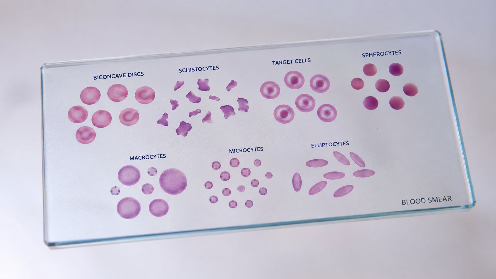
Schistocytes are fragmented red cells. They matter because fragmentation usually signals mechanical injury rather than a minor staining variation. Spherocytes are small, round cells that lack central pallor, which usually points toward membrane loss or membrane remodeling.
These two shapes can look very different under the microscope, yet both are informative because they suggest altered red cell survival. Schistocytes raise concern for shear injury or fragmentation, while spherocytes point toward loss of the normal biconcave shape. In practical interpretation, the morphology should still be matched with the clinical setting and the rest of the blood count, because the same appearance can carry different weight in different patients. For researchers building structured data sets, the OMOPHub guide for researchers on OMOP phenotypes is a practical resource for thinking about how morphology-related phenotypes are represented in analysis workflows.
Elongated, target, and spiculated cells
Target cells have a bullseye appearance, usually reflecting altered membrane-to-hemoglobin balance or surface area changes. Elliptocytes are oval or cigar-shaped cells, and they suggest a red cell that has lost its usual flexibility or structural symmetry. Spiculated cells, including echinocytes and acanthocytes, show surface projections that usually point to membrane or metabolic disturbance.
A quick visual rule helps here. Fragmented cells suggest breakage, rounded cells suggest membrane loss, elongated cells suggest distortion of the normal cell skeleton, and spiculated cells suggest a membrane surface problem. That framework is more useful than trying to memorize every named morphology in isolation, especially because laboratory preparation and slide quality can affect how strongly each feature appears. The same smear can look slightly different from one lab to another, so the pattern should be judged as a whole rather than by a single cell at the edge of the field.
Size changes still matter
Microcytes and macrocytes are size abnormalities rather than shape abnormalities, but they belong in the same mental map because size and shape often travel together. A small cell can reflect impaired hemoglobin synthesis, while a large cell can reflect altered maturation or membrane composition. On a smear, abnormal RBC morphology usually comes from both size and contour, not one alone.
That is why a report with mixed findings often needs careful reading. A population of smaller cells can make the smear look denser, while larger cells can seem more fragile or irregular even when the underlying problem is different. In the same way that a slightly bent coin and a flat coin are both still coins, altered red cells can share a visual category while arising from different biological problems.
Pathophysiological Mechanisms Behind RBC Shape Changes
Red cells keep their normal biconcave disc shape because membrane structure, hemoglobin content, and cytoskeletal flexibility work together. When one part of that system fails, the smear changes. The morphology then becomes a clue to the injured biological layer, not just the final shape seen under the microscope.
Membrane, hemoglobin, and marrow effects
Hemoglobin synthesis problems often alter cell size first, which is why microcytosis and some forms of macrocytosis belong in the same diagnostic conversation as shape abnormalities. Membrane protein and lipid defects can produce cells that are rounder, more fragile, or more irregular, because the cell no longer maintains its normal contour. Bone marrow dysfunction can also distort the output of circulating red cells, so a smear result has to be read alongside the broader hematologic picture.
The key idea is that morphology often reflects where the failure occurred. A membrane problem and a hemoglobin problem can both produce an abnormal smear, but they do not mean the same thing biologically. The same abnormal RBC morphology result can therefore lead to different workups depending on the associated CBC pattern and symptoms.
Mechanical and oxidative injury
Mechanical fragmentation produces schistocytes, because the cell is physically damaged as it moves through a hostile microenvironment. That is a different process from spiculation, where the cell membrane changes shape without being cut apart. In severe malaria, infected RBCs progressively lose their biconcave shape, become less deformable, and gain new cytoadherence properties, which helps explain microvascular obstruction and severity (malaria deformability review).
Oxidative or metabolic injury can also create spiculated cells. In hospitalized patients with COVID-19, spiculated cells were a prominent abnormality, and that pattern fits membrane protein and lipid disruption rather than a pure size disorder (COVID-19 RBC morphology study). As discussed later, COVID-19 studies also found spiculated cells to be the dominant change. The same principle helps explain why spiculated forms can show up in settings of renal, liver, or oxidative stress.
Clinical insight: if you see spiculation or fragmentation, think about membrane injury, oxidative stress, mechanical shear, and hemolysis together, not one at a time.
Diagnostic Approach to Interpreting Peripheral Smears
A smear becomes useful when it is read as part of a workflow. The first step is not naming the shape, it's confirming whether the shape fits the rest of the CBC and the clinical setting. That habit prevents overcalling incidental findings and helps you focus testing where it's most likely to pay off.

Start with the indices
Begin with hemoglobin, MCV, MCH, MCHC, and RDW. Those numbers tell you whether you are dealing with isolated shape change, mixed size abnormalities, or a broader anemia pattern. A morphology report that conflicts with a stable CBC deserves skepticism, or at least a careful second look at specimen quality and review method.
The same principle applies to the reticulocyte count. A smear suggesting hemolysis means more when the marrow response is raised, and less when the rest of the profile is flat. If the patient's symptoms, exam, and indices don't line up, the smear should prompt refinement, not automatic escalation.
Escalate testing by pattern
Additional tests should follow the morphologic pattern, not replace judgment. Iron studies fit microcytosis with supportive clinical context. Vitamin B12 and folate belong when macrocytosis or mixed indices raise concern for ineffective erythropoiesis. Hemoglobin electrophoresis makes sense when the smear and indices point toward a hemoglobinopathy. Hemolysis panels are most helpful when fragmentation, spherocytes, or other injury patterns suggest accelerated red cell destruction. Bone marrow evaluation becomes more relevant when morphology is persistent, unexplained, or paired with other cytopenias.
A molecular test can be helpful when the question is genetic, infectious, or clonally complex. For a broader overview of that testing mindset, the internal reference on molecular diagnostics is a useful companion piece.
Practical rule: order the next test that answers the most likely mechanism, not the test that sounds the most complete.
Clinical Significance in Infections and Systemic Disease
Abnormal red cell shape matters most when the patient is already sick. In that setting, the smear is not just a hematology artifact, it becomes a window into systemic stress, membrane injury, and microvascular disease. The challenge is learning which morphology is likely to be a marker of severity and which is just background noise.

Infection can reshape the smear
In hospitalized COVID-19 patients, 65% showed some degree of RBC morphologic abnormality at admission, and the most frequent change was spiculated cells (COVID-19 RBC morphology study). The same study reported a mortality gradient, with 41.9% mortality in patients with more frequent RBC morphological alterations, compared with 20.5% in those with fewer than 10% abnormal cells and 12.5% in those with no abnormalities, with p = 0.012 (COVID-19 RBC morphology study). That doesn't make morphology a standalone prognostic score, but it does show that a smear can carry meaningful clinical information during severe infection.
Malaria works through a different mechanism but points in the same direction. In Plasmodium falciparum infection, RBCs lose deformability and gain new cytoadherence properties, which helps drive microvascular obstruction and disease severity (malaria deformability review). The practical lesson is that morphology in infection often reflects the interaction between the parasite, the membrane, and the microcirculation.
Systemic illness can leave the same footprint
Spiculated cells aren't unique to one disease family. The COVID-19 study interpreted the finding as possible impairment of protein and lipid membrane composition (COVID-19 RBC morphology study), and that same membrane logic helps explain why liver, kidney, and oxidative stress states can produce abnormal forms. In other words, the smear may be telling you that the patient is under systemic pressure before the rest of the workup has fully declared why.
The internal discussion of laboratory diagnosis of viral infections is a useful adjacent read for clinicians who want to think more broadly about how infection severity shows up across routine testing.
Laboratory Reporting Variability and Interpretation Challenges
One of the least appreciated problems in smear interpretation is that two laboratories may describe the same specimen differently. That doesn't mean one report is wrong. It means reporting and grading practices vary across laboratories worldwide, despite advances in hematology analyzers and standardization efforts (Wiley hematology guidance).
Why the same smear can read differently
Manual smear review still depends on human judgment, especially when the abnormality is subtle or the population is mixed. Automated analyzer flags can suggest that something is off, but they don't always define the morphology with the same nuance a trained reviewer would use. The result is a report that may sound more alarming in one lab and more restrained in another.
That variability is exactly why a smear should be matched to the rest of the case. If the morphology seems inconsistent with the patient's clinical picture, review the specimen history, the CBC, and whether the finding came from automated flagging or manual confirmation. Differences in experience and local reporting conventions can change the wording without changing the underlying cells.
What to do when the report and the patient disagree
A good clinician doesn't argue with the lab, but also doesn't accept a discordant report at face value. Ask whether the finding was graded, how it was described, and whether a repeat smear would be more informative. If the result is important to management, a direct conversation with the laboratory is often the fastest way to resolve the discrepancy.
The broader lesson is that abnormal RBC morphology is a language with dialects. The cell doesn't change, but the report may. A careful reader learns to separate the morphology itself from the vocabulary used to describe it.
The internal guide on how to interpret lab results fits well here because the same interpretive discipline applies across many routine tests.
Practical Takeaways for Clinical Decision Making
A patient with mild poikilocytosis and otherwise normal indices is not the same as a critically ill patient with new spiculated cells. That sounds obvious, but the point gets lost when morphology is treated as a binary abnormal-or-normal switch. In real practice, the difference lies in context, persistence, and whether the smear agrees with the rest of the case.
Two common scenarios
A stable outpatient with a slightly irregular smear and reassuring CBC usually needs correlation, not panic. That means looking for nutritional issues, recent illness, specimen handling concerns, or a pattern that may be too mild to justify aggressive testing. If the patient feels well and the indices are clean, repeating the study or reviewing the original smear often makes more sense than launching a broad workup.
A hospitalized patient with newly spiculated cells is different. In that setting, the smear may reflect membrane injury, oxidative stress, or systemic illness, and the finding deserves prompt integration with hemolysis testing, renal and liver assessment, medication review, and the overall trajectory of the illness. When the morphology changes during acute disease, it should be treated as a clue to severity, not as an isolated laboratory curiosity.
A simple decision frame
Use three questions in sequence.
- Is the morphology substantial and reproducible? If not, verify the report.
- Do the CBC indices support the pattern? If not, reassess context.
- Does the clinical picture fit a membrane, hemoglobin, marrow, or hemolysis mechanism? If yes, choose the most targeted follow-up test.
Bottom line: abnormal RBC morphology deserves respect, but not reflexive alarm.
For teaching rounds, resident conferences, or bedside interpretation, keep the message tight, morphology is real, but it only becomes meaningful when it's placed next to the CBC, the smear quality, and the patient's story. If you want more clinician-focused explanations like this, visit VirusFAQ.com and use it as a practical teaching reference for your team, your students, or your next case review.
Leave a Reply