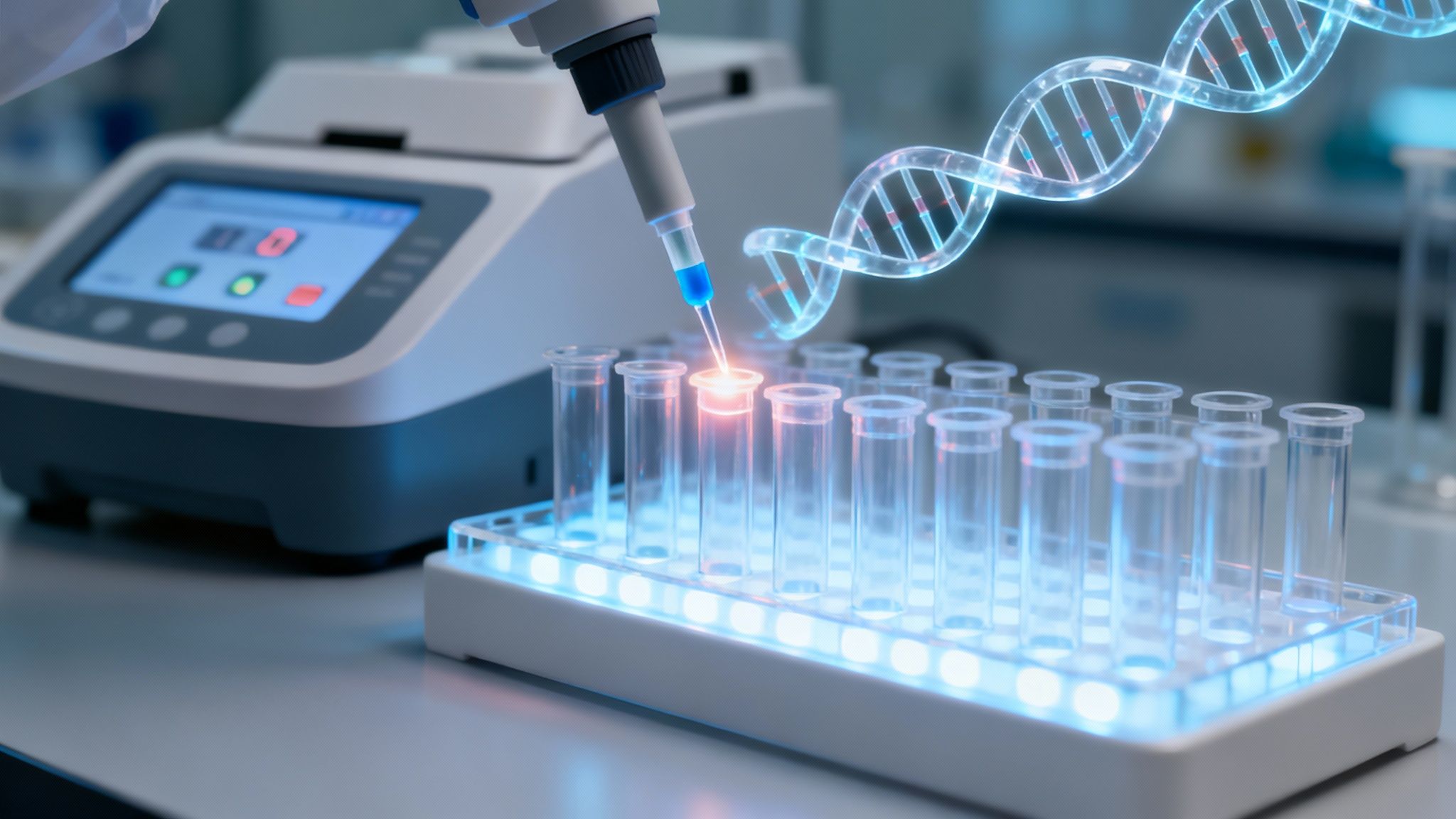
Polymerase chain reaction, or PCR, is basically a molecular photocopier. It gives scientists the power to take a tiny snippet of genetic material—think DNA or RNA—and rapidly make billions of copies.
This amplification is what makes it possible to spot specific genetic sequences, like those from viruses such as SARS-CoV-2 or Influenza, even when they're present in extremely small amounts. This is crucial for both diagnosis and for ensuring our environments are free from dangerous pathogens.
The Molecular Photocopier Behind Modern Diagnostics
Imagine trying to find one specific sentence hidden somewhere in the Library of Congress. It would be an impossible task. But what if you had a magical copier that could locate that single sentence and instantly print billions of copies, making it the most obvious thing in the room?
That’s exactly what Polymerase Chain Reaction (PCR) does for genetic material.
At its core, PCR is a method designed to make an exponential number of copies of a specific piece of DNA. This single capability is the foundation of modern diagnostics and a ton of other scientific breakthroughs. To really get how it works, we need to look at the key ingredients that make the whole process possible.
The Key Ingredients for Amplification
The PCR process is a bit like baking a cake—it requires a precise recipe of ingredients that work together in a machine that cycles through different temperatures. Each component has a critical job to do:
- DNA Template: This is your starting material. It’s the original sample that contains the specific genetic code you want to copy, like RNA from the Hepatitis C virus (HCV) that has been converted into DNA.
- Primers: Think of these as super-specific search terms. Primers are short, custom-built strands of DNA that find and latch onto the exact beginning and end points of the DNA segment you’re targeting.
- DNA Polymerase: This is the engine of your photocopier. It’s a special enzyme that reads the DNA template and starts building new, matching strands of DNA. It’s the real workhorse of the reaction.
- Nucleotides (dNTPs): These are the raw building blocks. If DNA polymerase is the engine, nucleotides are the "ink" it uses to construct all those new DNA strands.
When biochemist Kary Mullis invented this technique back in 1983, it was a massive leap forward for biology. His method of simply heating and cooling a sample could produce 100 billion copies of a DNA segment in just a few hours. It completely changed everything from forensic science to genetic research.
This powerful process is a cornerstone of what is molecular diagnostics, a field dedicated to analyzing the biological markers hidden in our genetic code. Understanding the science behind breakthroughs like this often starts with a good grasp of fundamental research methodology. Whether it's identifying pathogens like Norovirus and Human Rotavirus or helping solve a crime, PCR is what provides that clear, undeniable signal scientists need for an accurate diagnosis.
Decoding the Three Steps of a PCR Cycle
Polymerase chain reaction might sound intimidating, but it all boils down to a simple, repeating three-step cycle. Think of it like a highly controlled recipe where temperature is the key ingredient. By precisely raising and lowering the heat inside a thermal cycler, we can kick off a molecular chain reaction that turns a single strand of DNA into billions of copies.
The whole process relies on a few key players—the DNA template, primers, and a special enzyme called polymerase—all working together. This infographic gives a great visual of how they interact in each cycle.

As you can see, each component has a specific job, from finding the target DNA sequence to building the new copies. This teamwork is what drives the exponential amplification at the heart of PCR.
To get a clearer picture, let's break down what happens in each of the three stages of a single PCR cycle.
The table below gives a quick snapshot of the three core stages, their purpose, and the typical temperatures involved.
The Three Stages of a PCR Cycle
| Stage | Typical Temperature | Purpose |
|---|---|---|
| Denaturation | 94–98°C | To heat the sample and separate the double-stranded DNA into two single strands. |
| Annealing | 50–65°C | To cool the sample and allow primers to bind to their specific target sequences on the single DNA strands. |
| Extension | 72°C | To warm the sample to the optimal temperature for DNA polymerase to build new DNA strands, creating copies. |
Now, let's dive a little deeper into what each of those steps actually accomplishes.
Step 1 Denaturation
First, we crank up the heat. The cycle kicks off with denaturation, where the reaction is heated to a blistering 94–98°C. This intense heat is just enough to break the relatively weak hydrogen bonds holding the two strands of the DNA double helix together.
The result? Two single strands of DNA, now exposed and ready to serve as templates for the copying process. It’s a lot like unzipping a jacket to get to the fabric on both sides.
Step 2 Annealing
Next, we cool things down. The temperature is dropped significantly to somewhere between 50–65°C, which starts the annealing step. At this cooler temperature, the short, custom-designed DNA primers can zip around and find their exact matching sequences on the single-stranded DNA templates.
Once a primer finds its spot, it binds—or "anneals"—to it. This is probably the most critical step, because the primers act like signposts, telling the polymerase enzyme exactly where to start and stop copying. This ensures that only our target sequence gets amplified.
Key Takeaway: The incredible precision of PCR comes from the primers. Their unique sequence ensures that only the genetic material you care about—say, DNA from a specific virus like Hepatitis B Virus (HBV)—gets copied, while all the other DNA in the sample is ignored.
Step 3 Extension
For the final step, we turn the heat up again, but not as high as before. The temperature is raised to about 72°C, the perfect working temperature for our star enzyme, DNA polymerase. This stage is called extension (or sometimes elongation).
The polymerase latches onto the primer and starts zipping down the DNA template strand. As it moves, it grabs free-floating nucleotides from the mixture and adds them one by one to build a brand-new, complementary DNA strand. In an instant, a new double helix is formed.
Making this whole process practical and automated hinged on one major breakthrough: the discovery of Taq polymerase. This tough little enzyme was isolated from heat-loving bacteria in Yellowstone's hot springs. Unlike other enzymes, Taq can withstand the near-boiling temperatures of the denaturation step. This discovery was a game-changer, as it meant scientists no longer had to add fresh enzyme after every single cycle. You can explore the history of PCR to see just how much this transformed molecular biology.
At the end of this three-step cycle, we've doubled the amount of our target DNA. The machine then repeats this entire process another 30 to 40 times. With each new DNA strand becoming a template in the next round, the number of copies grows exponentially, turning a single molecule into billions.
Exploring Key Polymerase Chain Reaction Techniques
While the basic PCR cycle is a powerful tool, it’s really just the tip of the iceberg. Over the years, scientists have built on that foundation to create a whole toolkit of specialized polymerase chain reaction techniques, each designed to answer different and more complex biological questions. These variations take PCR from a simple "yes or no" detection method to a remarkably precise analytical instrument.
These advanced methods are the workhorses of modern diagnostics and research. They’re what let us not only find a virus but measure how much of it is there, detect viruses that use RNA instead of DNA, find incredibly rare genetic mutations, and even screen for multiple pathogens all at once.

Let's dive into four of the most significant PCR techniques that have become indispensable in labs worldwide.
Real-Time PCR (qPCR) For Counting Copies
Standard PCR tells you if your target DNA is present. But what if you need to know how much is there? That’s where Real-Time PCR, or quantitative PCR (qPCR), comes in. This technique doesn't wait until the reaction is over; it measures the DNA amplification as it happens, in real time.
Think of it like taking a photo after every single cycle of the molecular photocopier. By adding fluorescent dyes or probes to the mix, a qPCR machine detects the growing light signal as more and more DNA copies are made. The brighter the signal gets, the more DNA is present.
This allows scientists to work backward and figure out the exact starting amount of genetic material in the sample—a piece of data that's critical in many clinical settings.
Clinical Application: For patients with chronic viral infections like Hepatitis B Virus (HBV) or Human Immunodeficiency Virus (HIV-1), qPCR is used to measure their viral load. This tells doctors exactly how much virus is circulating in the body, which is vital information for tracking how well a treatment is working.
Reverse Transcription PCR (RT-PCR) For Detecting RNA Viruses
The central dogma of biology says that genetic information flows from DNA to RNA. But many viruses, including some of the most notorious pathogens out there, break this rule. Their entire genetic blueprint is made of RNA, not DNA.
This list includes viruses like:
- Influenza A Virus (H1N1)
- Hepatitis C Virus (HCV)
- SARS-Related Coronavirus 2 (SARS-CoV-2)
- Norovirus (Norwalk Virus) and Feline Calicivirus
Since standard PCR can only amplify DNA, it can’t directly "see" these RNA viruses. Reverse Transcription PCR (RT-PCR) solves this with a brilliant first step. Before the PCR cycles even start, an enzyme called reverse transcriptase converts the viral RNA into a stable, complementary DNA (cDNA) strand.
Once that conversion is done, the new cDNA acts as a perfect template for a standard PCR or qPCR reaction. This clever two-step process is the gold standard for diagnosing active infections caused by RNA viruses—it was the primary method used to detect SARS-CoV-2 during the global pandemic.
Digital PCR (dPCR) For Absolute Precision
What if you're looking for a needle in a haystack—say, a single mutated gene hidden among thousands of healthy ones? For jobs that demand the highest level of precision and sensitivity, scientists turn to Digital PCR (dPCR). This technique gives you an absolute, rather than relative, count of DNA molecules.
Instead of running one big reaction in a tube, dPCR partitions the sample into thousands of tiny, individual reactions. Imagine dividing a single drop of water into 20,000 microscopic droplets. Each droplet will contain either one or zero copies of the target DNA.
After the amplification cycles, the machine simply counts the "positive" droplets (the ones that glow) versus the "negative" ones. This binary, yes/no count provides a direct and stunningly accurate measurement of the target DNA's concentration, without needing a standard curve for comparison like qPCR does.
This makes dPCR incredibly useful for:
- Rare Mutation Detection: Identifying cancer-related mutations in a blood sample (a technique known as liquid biopsy).
- Copy Number Variation (CNV): Precisely measuring how many copies of a particular gene a person has.
- Low-Level Pathogen Detection: Finding trace amounts of viral DNA when an infection is in its earliest stages.
Multiplex PCR For High-Throughput Screening
In many clinical situations, a patient might have symptoms that could be caused by several different pathogens. Testing for each one individually would be slow, expensive, and a waste of time and resources. Multiplex PCR tackles this by detecting multiple genetic targets in a single reaction tube.
This is done by adding several different pairs of primers to the mix, with each pair engineered to find a different target. For example, a single Multiplex PCR test could simultaneously screen a respiratory sample for Influenza A, Influenza B, and Rhinovirus Type 14.
The efficiency is incredible. You save time, reagents, and precious sample material. It has become a cornerstone of syndromic testing panels in clinical labs, allowing for fast and broad screening for infectious diseases. The key is careful primer design to ensure they don't interfere with each other, but when it's done right, multiplexing dramatically speeds up the diagnostic workflow.
Choosing the Right PCR Method for the Job
With a few powerful polymerase chain reaction techniques in the lab's toolkit, picking the right one is like a mechanic grabbing the perfect tool for a specific engine repair. You wouldn't use a wrench to hammer a nail. In the same way, scientists have to match the PCR method to the exact question they need to answer—whether it’s simply identifying a virus, measuring how much of it is there, or hunting for a rare genetic marker.
The choice really boils down to what you need to measure and the level of precision required. Some tests just need a "yes or no" answer about a virus's presence, while others must answer "how much" is circulating in a sample. Getting a handle on these differences is the key to understanding why certain PCR methods are the gold standard for diagnosing and managing specific viral infections.
A Quick Look at the Major PCR Players
Each version of PCR has its own unique strengths. A qPCR test, for instance, is perfect for tracking how a chronic infection is progressing over time. On the other hand, RT-PCR is absolutely essential for diagnosing an active infection from an RNA-based virus like Influenza or Norovirus.
To make things clearer, let's break down the core functions and best-use cases for these lab workhorses.
Comparison of Major PCR Techniques
This table gives a side-by-side look at the most common PCR techniques, showing what they measure, what makes them special, and where you'll most often see them in action.
| Technique | Measures | Key Advantage | Common Application |
|---|---|---|---|
| qPCR (Quantitative PCR) | The quantity of DNA as it amplifies in real-time. | Provides a numerical value, allowing for precise quantification of the starting genetic material. | Monitoring the viral load of DNA viruses like Hepatitis B Virus (HBV) and HIV-1 to track treatment effectiveness. |
| RT-PCR (Reverse Transcription PCR) | The presence of RNA after converting it to DNA. | Enables the detection of RNA viruses, which are invisible to standard DNA-based PCR. | Diagnosing active infections from RNA viruses such as SARS-CoV-2, Hepatitis C Virus (HCV), and Rhinovirus Type 39. |
| dPCR (Digital PCR) | An absolute count of DNA molecules without needing a standard. | Delivers exceptionally high precision and sensitivity, ideal for detecting very rare sequences. | Identifying rare cancer mutations in blood samples or detecting low-level pathogens with absolute certainty. |
As you can see, each technique is finely tuned for a specific job. Now, let’s walk through a few practical scenarios to see how these choices play out in a real clinical setting.
Real-World Diagnostic Scenarios
For a doctor treating a patient with HIV-1, a simple yes-or-no result isn't enough. They need to know the patient's viral load—the exact amount of virus in the blood—to see if antiviral medications are working.
This is a perfect job for qPCR. By measuring the amount of viral genetic material over time, clinicians can make truly informed decisions about treatment. A falling number is great news, signaling the therapy is working. But if the number starts climbing, it could be a red flag for drug resistance. You can learn more about why these measurements are so critical in our guide explaining what is viral load testing.
Key Insight: The choice between PCR methods is driven by the clinical need. For chronic infections like HIV-1 or HBV, quantification (qPCR) is crucial for management. For acute infections like the Avian Influenza Virus (H5N1) or COVID-19, detection (RT-PCR) is the primary goal for diagnosis.
Now, picture a different scenario: a patient shows up with classic respiratory symptoms during flu season. The culprit could be an Influenza A virus (H1N1), a Rhinovirus, or even SARS-CoV-2. Since these are all RNA viruses, RT-PCR is the go-to tool. The test first converts any viral RNA into DNA and then amplifies it to give a clear positive or negative diagnosis, confirming an active infection.
Finally, let's step into a research lab. A scientist might be hunting for a rare drug-resistant viral mutation that shows up in only 0.01% of the viral population. In this case, neither qPCR nor RT-PCR might be sensitive enough to reliably spot it. This is where dPCR shines, giving researchers the absolute precision needed to count those few rare copies and help guide the development of new therapies.
How PCR Changed Everything in Health and Safety
What started as a niche lab technique has become a household name. Polymerase chain reaction (PCR) is more than just a scientific tool; it has completely reshaped modern medicine, public health, and even how we solve crimes. The simple but powerful idea of finding a single snippet of genetic code and making millions of copies has turned abstract science into a real-world force for good.
The most famous example, of course, is its role in diagnosing infectious diseases. PCR's incredible sensitivity and specificity make it the undisputed gold standard for sniffing out the genetic footprints of viruses and bacteria. This became crystal clear during the COVID-19 pandemic, when PCR tests became our primary weapon for tracking the spread of SARS-CoV-2 across the globe.
A Pillar of Infectious Disease Control
Long before 2020, PCR was the workhorse behind the scenes for managing viral outbreaks and chronic infections. It gives doctors the power to detect a huge range of pathogens with stunning accuracy, including:
- Human Immunodeficiency Virus Type 1 (HIV-1)
- Hepatitis B Virus (HBV) and Duck Hepatitis B Virus (DHBV)
- Herpes Simplex Virus 1 (HSV-1) and Herpes Simplex Virus 2 (HSV-2)
- Influenza viruses like H1N1, H2N2, and H5N1
The numbers tell the story. PCR can detect as few as 10 to 100 copies of a target DNA sequence in a sample. This allows for incredibly early disease detection and genetic screening with over 99% specificity. It’s no surprise the commercial market for PCR tools exploded into a multi-billion dollar industry, driven by these ever-expanding needs in the clinic.
Beyond the Doctor's Office
But PCR’s impact doesn't stop at the clinic door. In forensic science, it's become an essential tool for justice. Investigators can take a tiny, almost useless DNA sample from a crime scene—think a single hair or a speck of blood—and use PCR to generate enough genetic material for a DNA fingerprint. This has cracked countless cold cases and, just as importantly, freed many wrongly convicted people.
Back in the research lab, PCR helps scientists decode the secrets of our own DNA. By amplifying specific genes, they can pinpoint mutations tied to inherited diseases, watch how genes turn on and off to understand how our bodies function, and even develop new, highly targeted medicines.
The Power of Precision: Whether it's diagnosing a patient, solving a crime, or verifying the cleanliness of a surface, every PCR application lives and dies by one thing: precision. Just one stray molecule of contaminating DNA can be amplified by mistake, leading to a completely wrong result. This is why a sterile, meticulously clean lab environment isn't just a recommendation—it's an absolute necessity.
The constant push to make molecular diagnostics like PCR even better is often powered by dedicated research funding. For example, recent initiatives providing grant funding to transform sarcoidosis research help drive the innovation that makes these tools faster, cheaper, and more powerful.
At the end of the day, whether it’s in a cutting-edge research institute or a small local clinic, the goal is the same: accurate, reliable detection. To see how PCR fits into the bigger picture, take a look at our guide on the laboratory diagnosis of viral infections.
Troubleshooting Common PCR Pitfalls
For all its power, a successful PCR experiment lives and dies by precision. We’re talking about a technique designed to amplify the tiniest traces of genetic material, so it’s no surprise that even the smallest mistake can lead to confusing—or just plain wrong—results. Staring at a failed or ambiguous experiment is a rite of passage for every scientist, but understanding the likely culprits is the first step to fixing the problem.
Most of the time, troubleshooting begins with a long, hard look at your gel electrophoresis results. In a perfect world, you’d see a single, bright, beautiful band right where it’s supposed to be. But reality often serves up frustrating issues like faint bands, a bunch of extra bands, or—the worst—no bands at all.

Each of these outcomes is a clue, pointing you toward a different potential problem, from wonky ingredients to bad temperature settings.
Identifying No Amplification Signal
Seeing a completely blank gel is one of the most common—and soul-crushing—PCR problems. This total lack of a signal usually means a critical component of the reaction either failed spectacularly or was missing from the get-go.
Before you throw everything out and start over, it’s worth checking a few simple things:
- Reagent Integrity: Are you sure you added everything? It’s surprisingly easy to forget a key ingredient like the DNA polymerase or primers. Also, make sure all your reagents were properly thawed and mixed before you started.
- Template Quality: Your starting DNA might be degraded or, worse, contain inhibitors that are killing the reaction. Poor sample quality is a notorious amplifier of failure.
- Incorrect Annealing Temperature: If the temperature is too high, your primers can’t grab onto the DNA template. If they can’t bind, the reaction never even starts.
Dealing with Non-Specific Bands and Primer-Dimers
Then there's the opposite problem: you get too much of a good thing. Seeing multiple unexpected bands, known as non-specific amplification, is a sign that your primers are getting a little too friendly and binding to the wrong DNA sequences. This often happens when the annealing temperature is too low, giving the primers a chance to stick where they shouldn’t.
A related headache is the appearance of a tiny, faint band way down at the bottom of the gel. This little guy is often a primer-dimer, which happens when the primers decide to bind to each other instead of doing their job and finding the DNA template.
Crucial Consideration: One of the sneakiest and most significant causes of failed or inaccurate PCR results is environmental contamination. Since you're amplifying traces of viral DNA, even a single stray skin cell or a contaminated pipette tip can introduce foreign genetic material into your reaction, leading to false positives and confusing results.
This is exactly why a sterile workspace isn't just a suggestion—it's non-negotiable. Regularly disinfecting your surfaces and equipment with quality wipes isn't just good lab practice; it’s essential for protecting the integrity of your results. Every single step must be taken to ensure the only DNA you amplify is the DNA you actually want.
Your PCR Questions, Answered
As you get more familiar with polymerase chain reaction, a few common questions always seem to pop up. Let's clear up some of the most frequent ones to help you lock in your understanding of this incredible technology.
Just How Sensitive Is a PCR Test?
PCR is exceptionally sensitive. We're talking about the ability to reliably spot as few as 10-100 copies of a specific DNA or RNA sequence hiding in a sample.
This incredible level of detection is precisely why it’s the gold standard for catching viral infections like HIV-1 or Influenza early on, when the pathogen's genetic footprint might be almost undetectable by other means.
What’s the Real Difference Between PCR and qPCR?
The main distinction comes down to one thing: measurement.
Think of a standard PCR test as a simple yes/no question—it tells you if your target DNA sequence is present at the end of the run. In contrast, quantitative PCR (qPCR) is all about the numbers. It measures the DNA amplification in real-time, cycle by cycle, letting you figure out the starting amount of the DNA or RNA you were looking for.
Why Does Everyone Stress a Clean Environment for PCR?
The extreme sensitivity that makes PCR so powerful is also its greatest weakness.
Because PCR is designed to amplify minuscule amounts of DNA, it is highly vulnerable to contamination. Even a single stray DNA molecule from another source—like a skin cell, a different sample, or dust—can be amplified, leading to a false-positive result.
Even one rogue DNA molecule floating in from dust, a skin cell, or a previous experiment can get amplified, creating a false positive and completely ruining your results.
That's why a sterile work area is non-negotiable. Strict disinfection routines, including the use of high-quality surface wipes, and dedicated equipment are absolutely essential to make sure the only DNA you’re detecting is the DNA from your sample. It's the only way to guarantee your results are accurate and trustworthy.
Leave a Reply