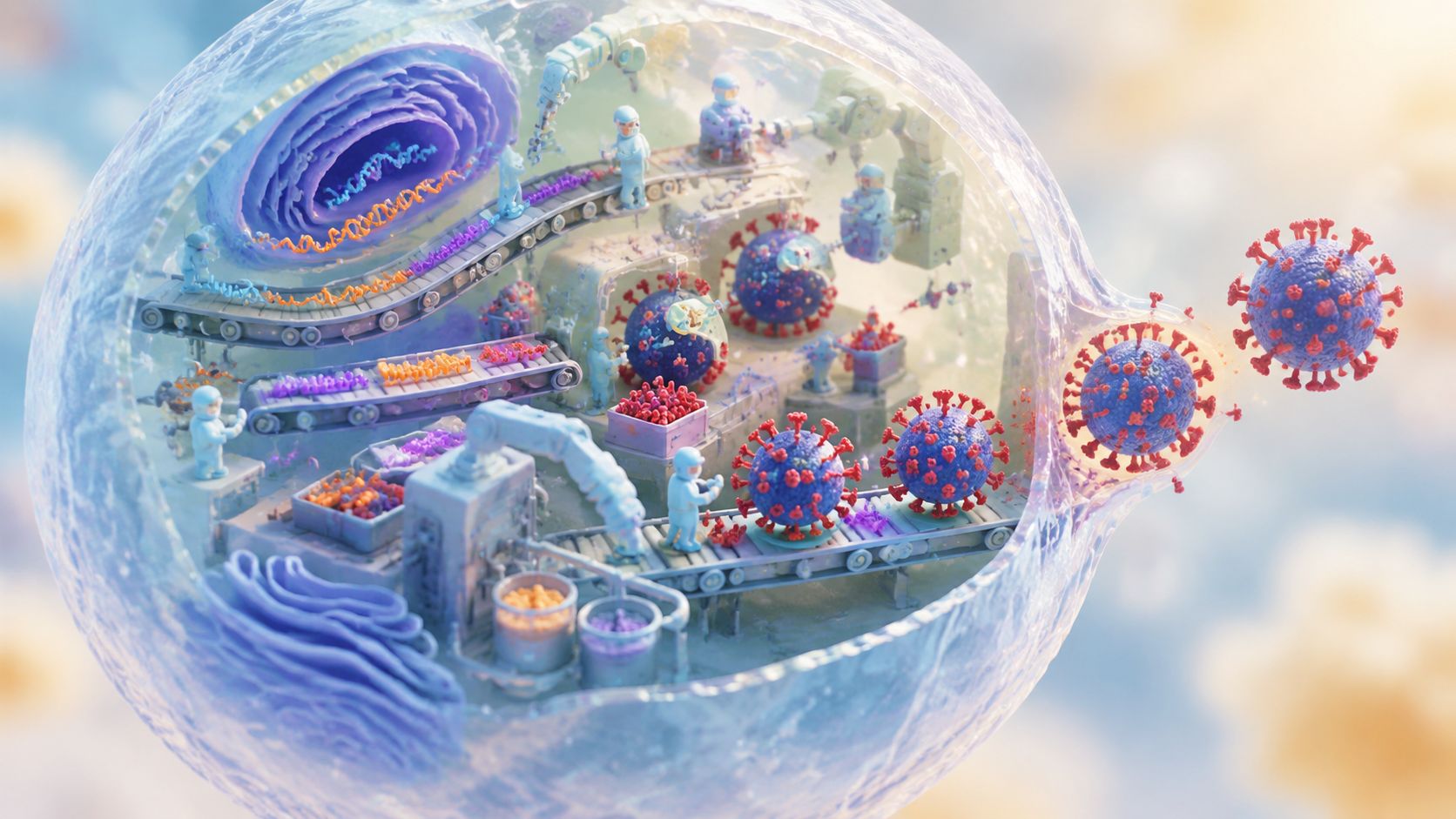
Influenza A can complete its first productive cycle and shed newly formed viruses from an infected cell in as little as 6 hours after attachment, internalization, and genome release (review of influenza replication). That speed helps explain why a respiratory infection can expand before symptoms fully reveal what's happening.
The influenza virus life cycle isn't just a straight line from entry to budding. It's a tightly coordinated operation involving viral proteins, host-cell machinery, changing endosomal acidity, nuclear transport, and a key release step. Many exposures never produce a successful infection because the virus must pass several intracellular bottlenecks, not merely attach to a cell.
This guide follows the process in order, beginning with the virus's structure and receptor preferences, then moving through entry, uncoating, nuclear transcription, genome replication, assembly, budding, and release. It also examines the less visible host controls that determine whether an incoming particle succeeds, and why interrupting transmission outside the cell still matters.
How the Influenza Virus Life Cycle Unfolds in Hours
Someone inhales respiratory particles containing influenza A. Some virions encounter the right type of airway cell, while others are trapped, cleared, or fail to begin productive replication. For a successful particle, the first contact is only the opening move in a compressed cellular production schedule.
The virus attaches to sialylated receptors on the cell surface through hemagglutinin. The cell then draws the particle inward inside an endosome, a membrane-bound compartment that acts like a temporary transport chamber. As the chamber acidifies, the virus undergoes carefully timed molecular changes that expose its genome and prepare it for the nucleus.
A short intracellular production line
Influenza A's first new viruses can be shed in as little as 6 hours (influenza replication review). That timeline compresses several jobs into one cycle:
- Attachment and uptake, where hemagglutinin recognizes a compatible receptor and the cell internalizes the virion.
- Uncoating, where acid-triggered changes loosen the viral package.
- Nuclear import, because influenza's ribonucleoprotein complexes must reach the nucleus.
- Transcription, including cap snatching from host RNA.
- Genome replication, which creates new viral RNA segments.
- Protein production, assembly, budding, and neuraminidase-driven release.
The analogy of a factory is useful, but it has limits. A factory usually receives raw materials from outside. Influenza brings its own genetic instructions, then redirects the host cell's equipment to manufacture viral parts while also avoiding cellular defenses.
Central idea: Entry starts infection, but productive infection requires the virus to pass every major checkpoint that follows.
Why this biology matters
For general readers, the life cycle explains why influenza can spread rapidly through respiratory tissue and why prevention must interrupt transmission before particles reach another cell. For students and specialists, it highlights intervention points such as the M2 proton channel, viral polymerase, nuclear export, and neuraminidase.
The sections ahead move from the virus's physical design to its timed entry mechanism, then into the nucleus where viral RNA is transcribed and copied. The final stages show how components return to the cell surface, form new particles, and escape. A separate discussion then examines host modifications and the early transcription bottleneck that can stop an infection even after entry.
What Makes Influenza Viruses Tick
Influenza A is an enveloped RNA virus. Its outer lipid membrane carries proteins that help it attach to cells, enter them, and leave after replication. Inside, segmented viral RNA is wrapped with proteins into viral ribonucleoproteins, or vRNPs, which function as protected genetic cargo.
A simple lock-and-key analogy helps:
- Hemagglutinin, or HA, is the attachment key. It recognizes sialic-acid-containing structures on host cells and later helps membranes fuse.
- Neuraminidase, or NA, is the release tool. It cuts terminal sialic acid residues so newly formed particles can escape rather than remain stuck.
- M2 is an ion channel in the viral envelope. It helps acid-triggered uncoating by allowing protons into the virion.
- M1 is a matrix protein beneath the envelope. It supports particle structure and later assists vRNP movement and assembly.
- vRNPs are the genome packages. They carry viral RNA together with nucleoprotein and polymerase components.

Receptors shape tissue preference
The key isn't whether a cell has sialic acid. The chemical linkage and presentation of that receptor influence whether HA can bind efficiently. This preference contributes to viral tropism, meaning the tissues and cell types a virus can infect effectively.
Tropism is a compatibility problem. A virion may reach a tissue, but successful infection depends on whether its attachment protein, entry process, and intracellular requirements match the cells it encounters.
That compatibility helps explain why influenza strains can behave differently in the respiratory tract. Some receptor preferences favor infection in regions where particles transmit efficiently, while others can support infection deeper in the lungs, where disease may be more severe. Receptor binding is important, but it doesn't guarantee success. The virus still needs to uncoat, transcribe its genome, evade defenses, assemble, and release.
Readers who want a broader introduction to viral genetic material can explore what RNA viruses are. Influenza is especially instructive because its RNA genome operates through a nuclear, segmented replication strategy rather than a simple cytoplasmic process.
How Influenza Enters Cells and Uncoats Its Genome
Influenza entry works like a timed lock with more than one setting. The virus first attaches to a compatible sialylated receptor, then the host cell encloses it through receptor-mediated endocytosis. The resulting endosome carries the virion inward and gradually changes its acidity.
That acidification matters because influenza uses different pH conditions to activate different molecular events. The early change helps prepare the internal viral machinery, while the later, stronger change triggers membrane fusion and deeper uncoating.
The first acid trigger
As the endosome acidifies to about pH 6, the viral M2 proton channel becomes active (influenza entry and uncoating review). Protons move into the virion, changing its internal environment and weakening interactions that hold the viral package together.
M2 doesn't fuse the viral and endosomal membranes. Instead, it helps create the conditions for uncoating. That distinction prevents a common confusion: M2 controls proton flow, while HA performs the membrane-fusion role after the appropriate low-pH trigger.
The second acid trigger
The endosome later reaches roughly pH 5.0 to 5.5. At this stage, M1 dissociates from the vRNPs, and HA undergoes an irreversible conformational change (mechanistic entry review). HA then drives fusion between the viral envelope and the endosomal membrane.
Fusion opens a route for the vRNPs to leave the endosomal compartment. The viral genome isn't yet translating proteins in the cytoplasm. Instead, the vRNPs must travel toward and enter the nucleus, where influenza carries out transcription and genome replication.

The pH dependence creates a potential vulnerability because the virus needs the sequence to occur in the correct location and order. If acidification, M2 function, HA activation, or membrane fusion fails, the genome remains trapped or cannot reach the machinery needed for productive infection.
For a broader explanation of attachment, penetration, uncoating, replication, assembly, and release, see how viruses infect cells. Influenza adds a distinctive layer because its genome enters the nucleus and depends on a specialized polymerase strategy there.
Transcription Replication and Protein Production Inside the Nucleus
Once vRNPs reach the nucleus, influenza begins converting its genetic information into usable messages and new genome segments. This is one of the virus's most distinctive features. Rather than relying on a conventional cellular mRNA start, its polymerase performs cap snatching.
The viral polymerase binds host RNA polymerase II and an emerging host transcript. It then cleaves off a capped 5′ RNA fragment and uses that fragment to prime viral mRNA synthesis (study of influenza cap snatching). The stolen cap helps the viral message remain stable, leave the nucleus, and engage host ribosomes for translation.
Hijacking the host printing press
Think of the nucleus as a publishing room. Influenza brings the template, but it borrows the host's formatting equipment. Cap snatching gives viral mRNAs a recognizable beginning, allowing host-cell systems to treat them as messages suitable for protein production.
The viral polymerase produces mRNAs that move out of the nucleus to the cytoplasm. Ribosomes then translate those messages into proteins such as HA, NA, M1, and NP. HA and NA are directed toward cellular membranes, while internal proteins help package the genome and coordinate later stages.
Making messages and making genomes
Transcription and genome replication are related but distinct jobs. Transcription creates mRNAs, which are readable instructions for protein production. Replication creates new copies of the viral RNA genome, including the segments that will be packaged into progeny vRNPs.
The polymerase uses complementary RNA as an intermediate during genome copying. New vRNA segments associate with nucleoprotein and polymerase components, producing vRNPs that can eventually leave the nucleus.

This division of labor creates another checkpoint. The cell must produce enough viral proteins, the polymerase must copy the genome correctly, and the new segments must remain associated with the right packaging machinery. A successful infection therefore depends on coordination, not solely on the presence of viral RNA.
The result is a supply of structural proteins and newly formed vRNPs. Those parts still occupy different cellular locations, so influenza must organize their movement before complete particles can form.
Assembly Budding and Release of New Influenza Viruses
The final part of the influenza virus life cycle resembles a carefully managed exit operation. New vRNPs must leave the nucleus, viral proteins must reach the appropriate region of the cell membrane, and the components must gather into a particle with the correct outer proteins and genome cargo.
Nuclear export and trafficking
Influenza's nuclear export factor, NEP, is described as the minimal essential viral factor required for vRNP nuclear export. Matrix protein M1 makes export more efficient and helps influence vRNP localization after the complexes reach the cytoplasm (research on influenza vRNP export).
After export, vRNPs move toward the plasma membrane. HA, NA, and M2 also accumulate in membrane regions that support particle formation. M1 acts as an organizing layer between the viral envelope and internal cargo, helping connect the genome packages with the budding membrane.
Budding isn't the same as release
The membrane bends around the assembled components and pinches outward, creating a new enveloped virion. But budding alone doesn't guarantee escape. Newly formed particles can remain attached to sialic-acid-containing structures on the host surface or bind to one another.
NA solves that problem by cleaving terminal sialic acid residues from cellular glycoconjugates and viral particles. This prevents self-aggregation and reduces the chance that a progeny virion will attach back to the cell it just left (review of influenza release).
Practical interpretation: HA helps influenza attach, while NA helps newly formed particles detach. Efficient spread depends on the balance between those opposing activities.
This is why NA is a validated antiviral target. Blocking its activity can leave particles formed but poorly released, limiting onward dissemination even when earlier replication steps have occurred.
Hidden Host Controls and Why Most Infections Fail
The textbook version of influenza replication often looks linear. In reality, host-cell regulation affects several stages at once. A 2026 review describes phosphorylation, ubiquitination, SUMOylation, glycosylation, acetylation, lipidation, and RNA methylation as a coordinated network influencing entry, replication, immune evasion, and assembly (2026 review of host post-translational modifications).
These modifications act like molecular switches, labels, and routing signals. They can change the activity, stability, location, or interaction partners of viral and host proteins. That means a target that looks essential in one stage may behave differently when the entire cycle is considered.
The early transcription bottleneck
Single-virus tracking in a 2026 study reported that only a minority of influenza infections succeed and identified viral transcription as the largest bottleneck (Hubrecht Institute summary of the single-virus study). Failures in the earliest viral RNA readout sharply reduce the chance that the infection becomes productive.
This finding changes the usual emphasis. Receptor binding and entry are necessary, but they aren't sufficient. A virus can enter a cell and still fail if its genome doesn't produce an effective early transcription signal.
The crucial question isn't only whether influenza gets inside. It's whether the incoming genome starts working quickly enough to establish control.
Host defenses add another layer. Cells detect viral activity, alter signaling pathways, and deploy antiviral responses. Readers seeking a focused explanation of this response can review how interferon works. The outcome depends on timing, viral protein function, host modifications, and the quality of the first transcriptional events.
This broader view explains why antiviral development is difficult. A compound may block one step while the virus uses compensatory interactions elsewhere. Effective strategies must account for the connected network rather than treating entry, replication, immune evasion, and assembly as isolated modules.
What the Life Cycle Teaches Us About Stopping Influenza
Influenza's rapid biology gives prevention a clear logic. Because newly formed virus can be shed from an infected cell in as little as 6 hours (influenza replication review), reducing exposure before particles reach airway cells remains valuable. Ventilation, staying home when ill, covering coughs, and hand hygiene all reduce opportunities for infectious material to move from one person or surface to another.
The same core cycle applies across influenza A viruses, including H1N1, H2N2, and H5N1, although receptor preferences, tissue tropism, host range, and disease behavior can differ. HA still supports attachment and fusion, M2 still participates in acid-triggered uncoating, the polymerase still handles viral RNA synthesis, and NA still supports efficient release.
Practical interruption points
- Reduce contact with respiratory particles. Improve airflow where people gather and avoid close contact during active illness.
- Clean frequently touched surfaces. Use an appropriate disinfecting product, follow its label directions, and respect the required contact time. Disinfecting wipes can make targeted cleaning practical for handles, switches, phones, and shared work surfaces.
- Protect your hands. Wash with soap and water when available, especially before touching your eyes, nose, or mouth.
- Use reliable education. VirusFAQ.com provides educational and scientific articles about viral biology, transmission, and prevention, including explanations of infection stages and disinfection.
Cleaning doesn't enter an infected cell or block polymerase activity. Its role comes earlier, by reducing the chance that virus-containing material remains available for transfer. Understanding the life cycle makes that distinction clear: prevention succeeds when it interrupts the chain before attachment begins.
Choose a disinfecting wipe that is labeled for the surfaces you need to clean, keep it accessible in high-contact areas, and use it consistently alongside ventilation, hand hygiene, and sensible illness precautions.
Leave a Reply